Do you experience any of the following symptoms after receiving a strong impact on your eyes?
- When you look at it with both eyes, it looks like a duplicate
- My eyes are sunken
- My eyes have become smaller
The cause of enophthalmos is Orbital Fracture, Enophthalmos after inadequate fracture repair and Congenital exophthalmos (by nature)
目次
The orbit is the space that contains the eyeball.
The inside and bottom of the orbital wall at the back of the eye socket is made of thin bone, so if the eye receives a strong impact, it may break.
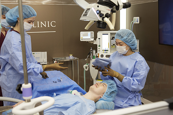
This is called an orbital blowout fracture. At our hospital, we perform orbital fracture surgery by approaching from the white of the eye (conjunctiva) without cutting the skin.
Although a high degree of technical skill is required, by approaching from the white of the eye (conjunctiva), it is possible to reduce the amount of post-operative damage in a short period of time.
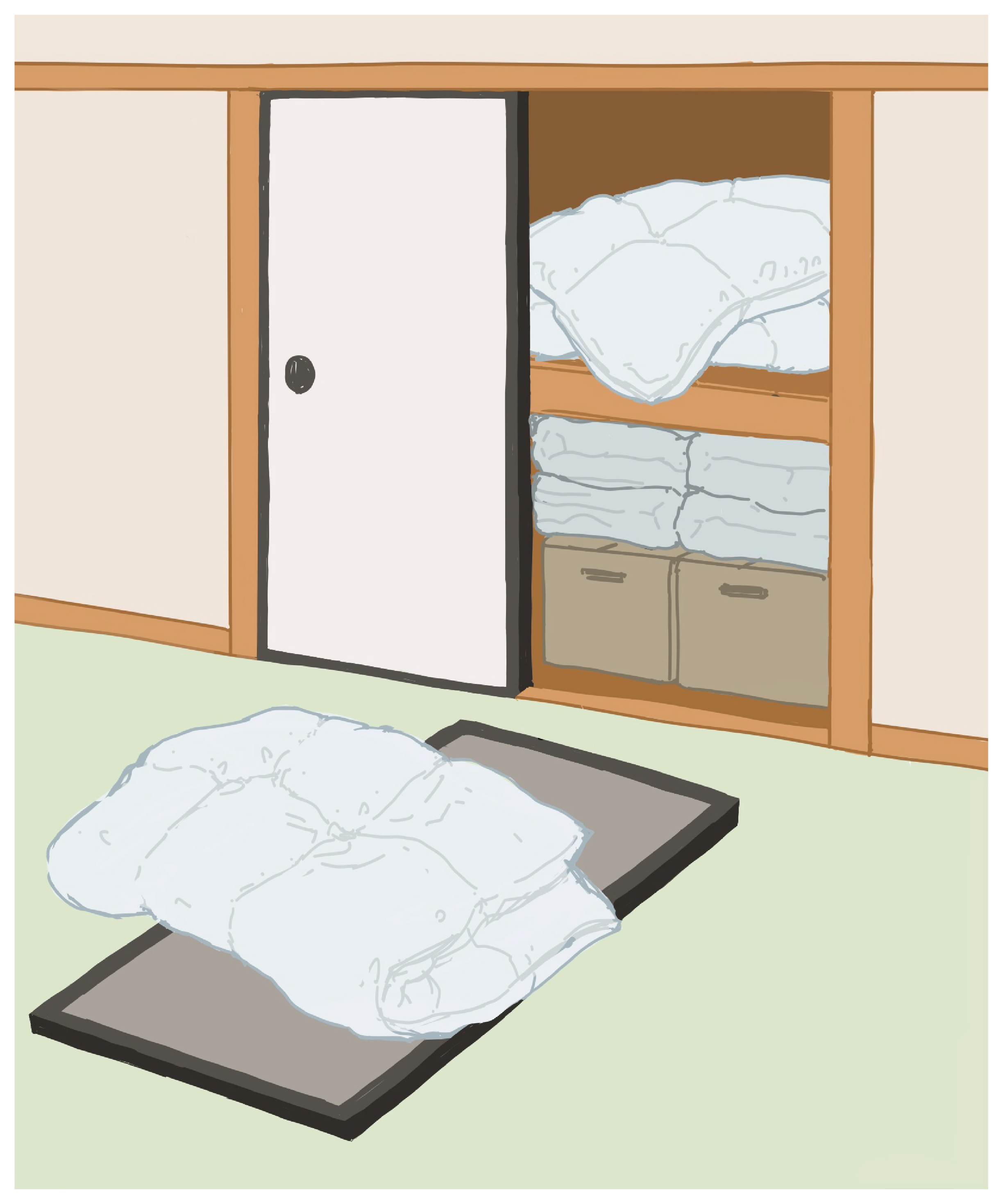
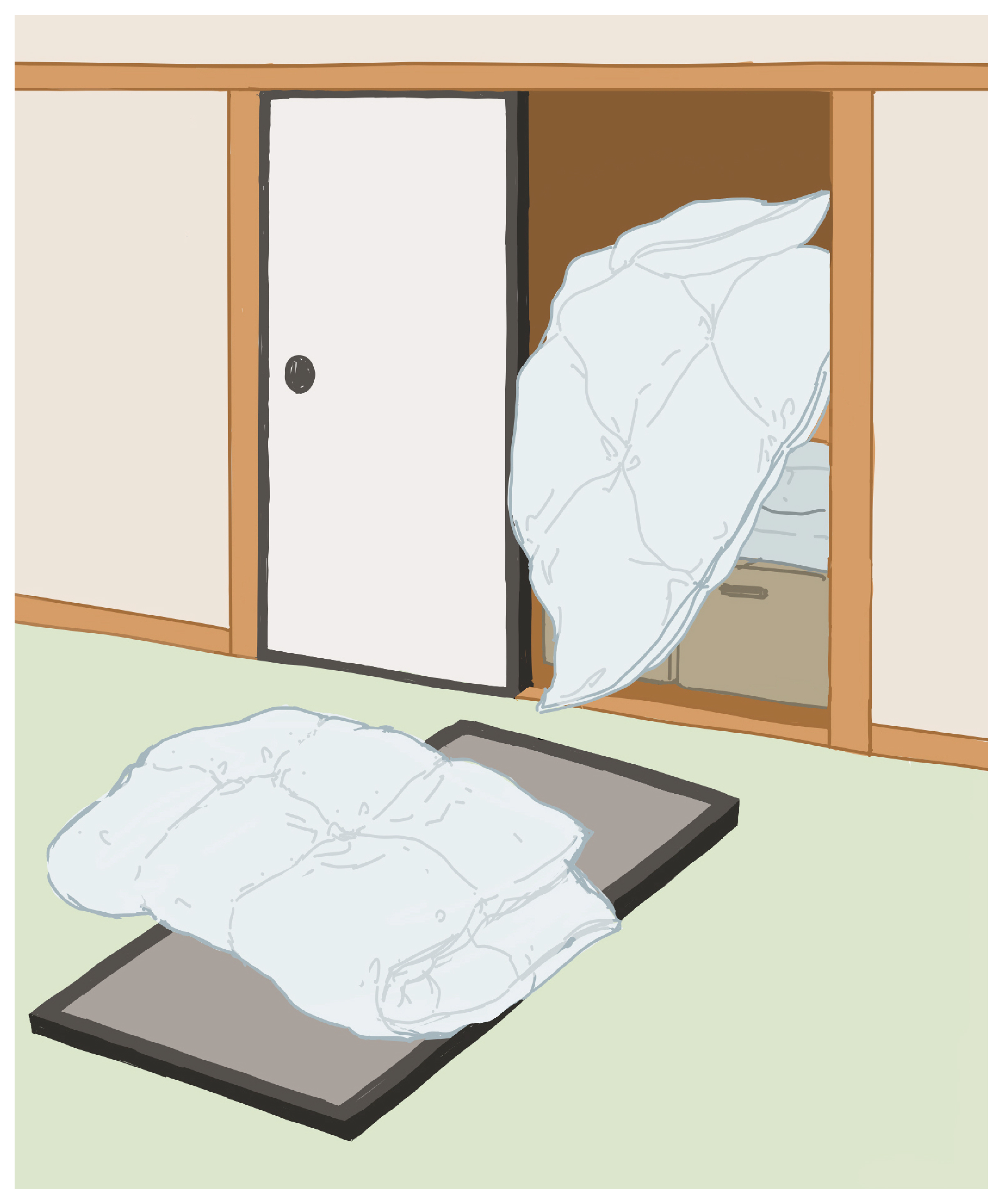
The cause of these injuries is when the eye receives a shock, which causes the bones deep around the eye to break, causing tissues such as muscles and fat behind the eye to shift. This can be compared to a situation where a futon is neatly placed in a closet, but in the case of an orbital fracture, the sliding door falls down and the futon is sticking out.
If the muscles are pulled at this time, it will cause the eyes to look double, and if the tissue behind the eyes, such as fat, prolapses, the eyes will become sunken and smaller. This is the main symptom of orbital fracture.
Additionally, if a fissure fracture occurs and tissue becomes trapped in the area, it becomes a closed fracture, which impairs blood circulation and causes tissue necrosis, necessitating early surgery.
1.Normal
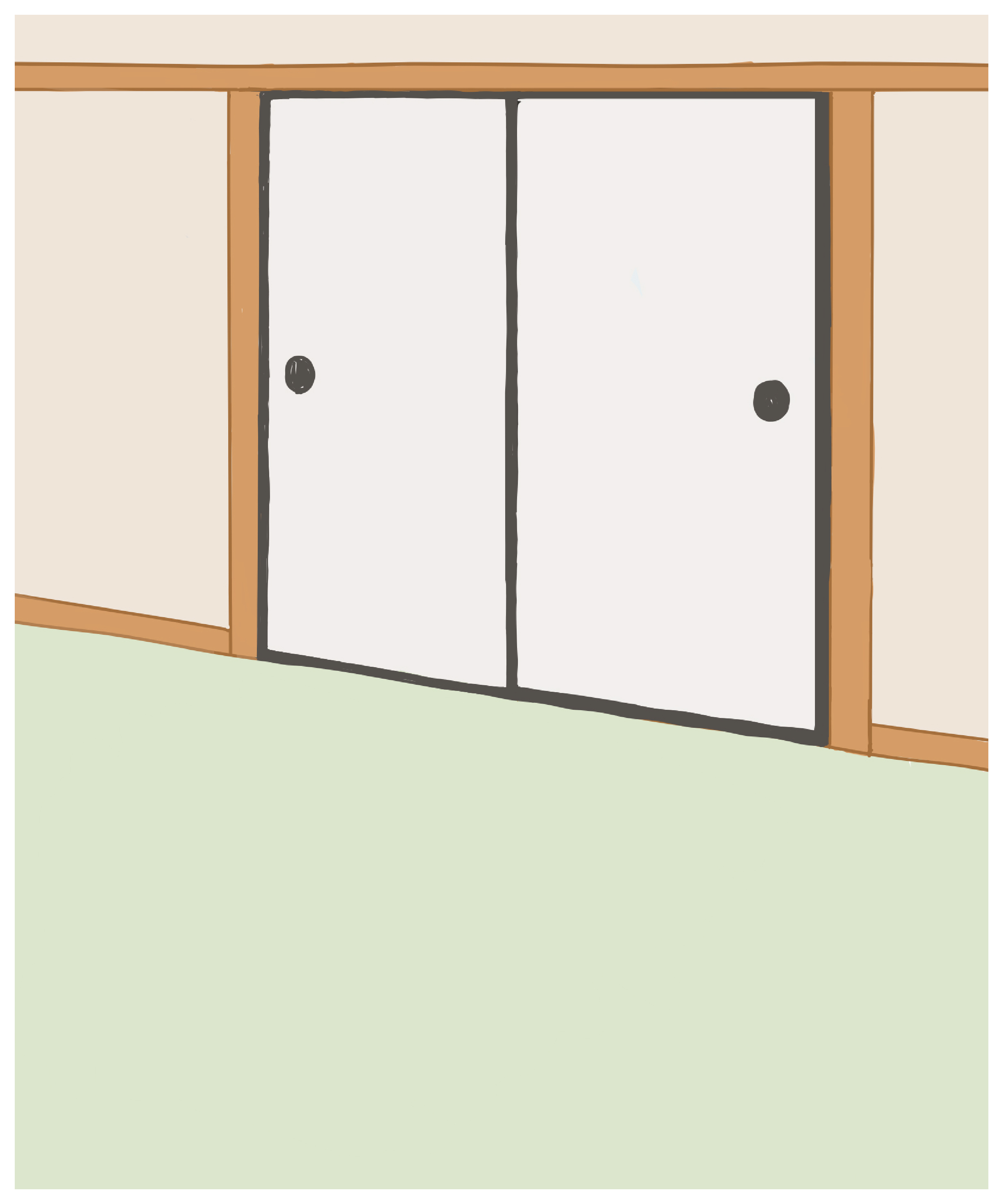
2.Neatly organized
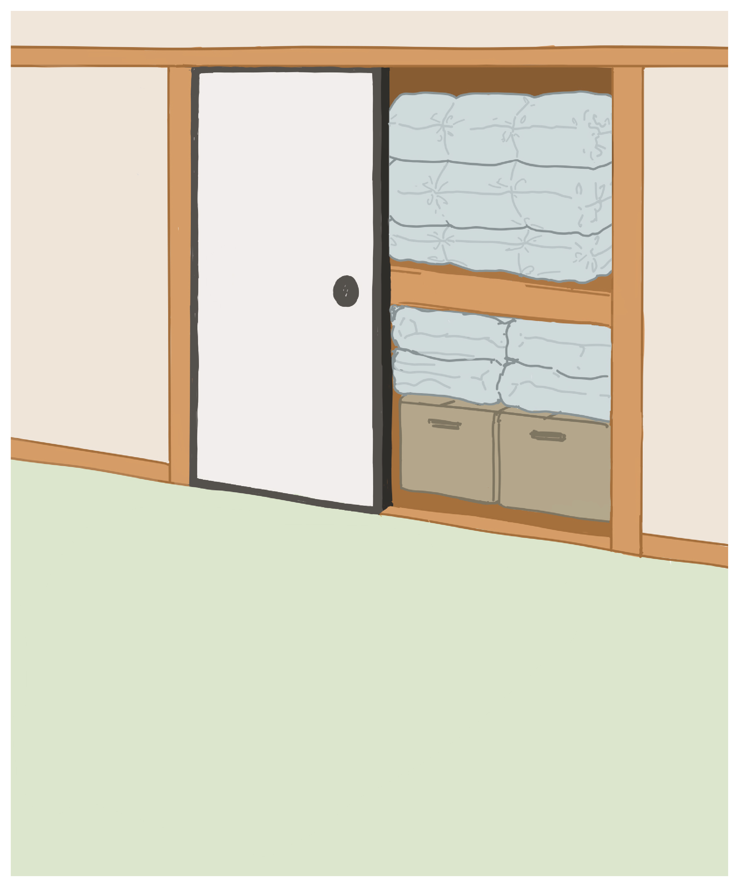
3.The futon flies out
4.Orbital fracture condition
5.Treatment of orbital fractures
Treatment involves returning tissues such as fat and muscle that have moved to the fracture site to their original positions.
However, if you simply return the fat and muscle to their natural position, they will return to that space.
Therefore, either the broken bone is removed and returned to its natural position, or an artificial bone is used in its place to return it to its natural position.
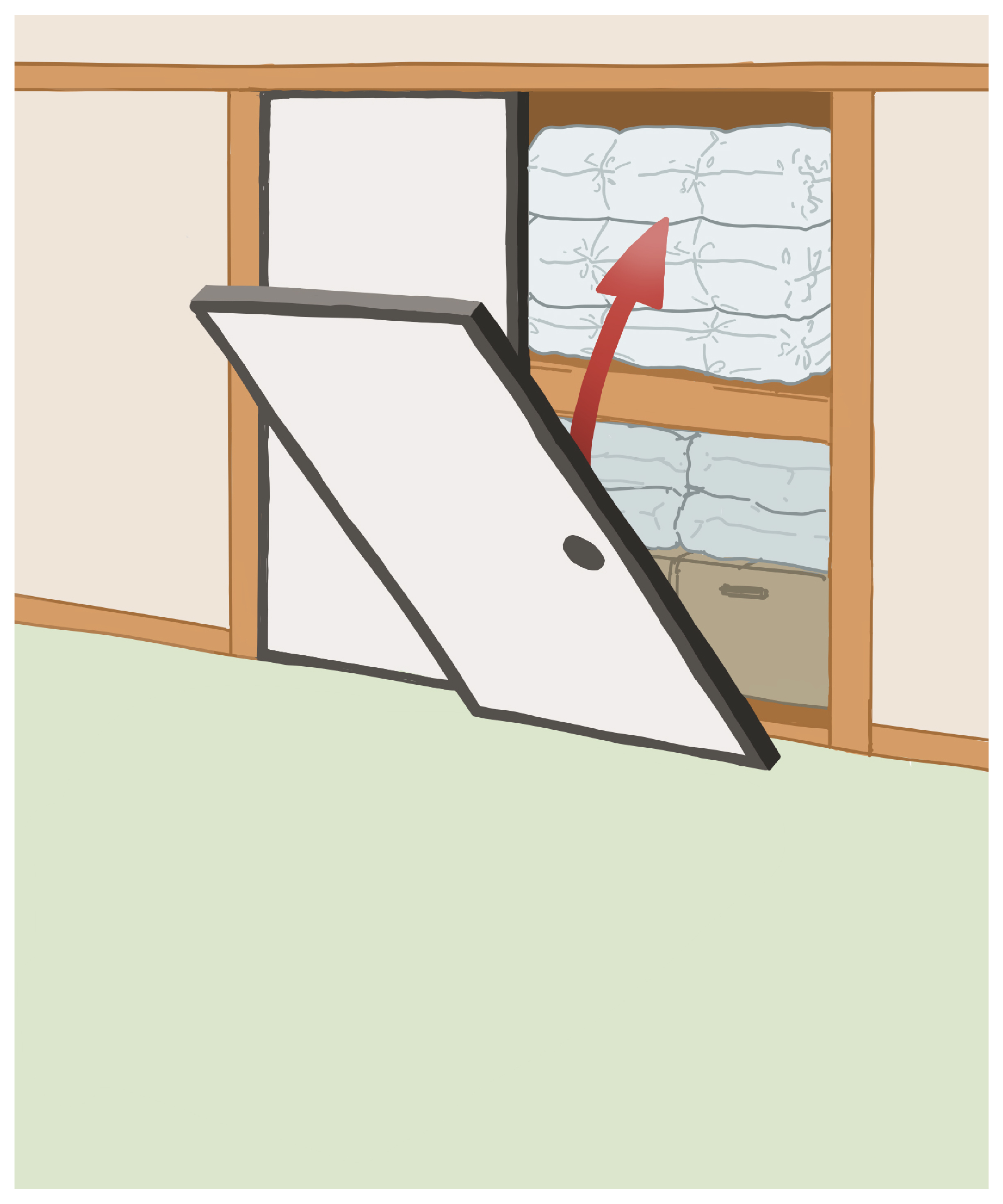
If we were to use the analogy of a closet, we would first put the futon that had fallen out into the closet, then put the fusuma back in its original position.
In the case of a closed fracture, the symptoms are often severe and there is a high possibility of residual effects, so it is necessary to undergo surgery by the next day.
It is recommended that surgery be performed within 24 hours if possible.
As mentioned earlier, treatment involves returning the displaced tissue to its original position and rebuilding the bone to prevent it from shifting again.
However, even if the tissue is returned to its original position, if the bone cannot be rebuilt to its original position, the tissue will move again.
If we were to use the analogy of a closet, even if you were able to put the futon back in its original position, if you couldn't get the fusuma into the correct position, the futon would come out again.
If the healing process is insufficient despite surgery at another hospital, there is a possibility that the bone reconstruction was not successful.
Using the analogy of a closet, the fusuma must be rebuilt in its original position, but there are some cases where a new fusuma is placed on top of the fallen one and that's it.
In this case, the fusuma has not healed properly, so the futon will come out again.
At our hospital, we also perform re-surgery for patients whose condition did not heal or worsened after surgery at another hospital. Please feel free to contact us.
At our hospital, orbital fracture surgery is approached from the white of the eye (conjunctiva), so no scars are left on the skin.
This is the world's most advanced surgical method that I learned from studying abroad in the United States. By not cutting the skin, not only does it leave no scars, but it also shortens the surgery time and reduces the burden on the body.
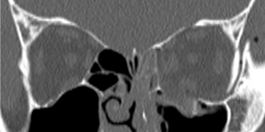
The space surrounded by bones containing the eye is called the orbit, and the bone surrounding the eye socket is called the orbital bone. The lower part is the maxillary sinus, and the inner part is the ethmoid sinus, which is a small space.

The inner wall is almost flat. The space behind the inner wall contains the ethmoid sinus.
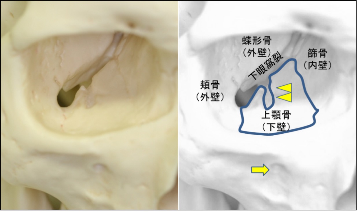
A large fissure (inferior orbital fissure) is visible in the center of the inferior orbital wall. In the space behind the lower wall is the maxillary sinus.
When you hit your eye, these bones break as they absorb the impact.
At the same time, orbital tissue (fat and muscle) protrudes into those spaces, a condition known as an orbital fracture.
Depending on where it breaks, it is said to be an inferior orbital wall fracture, an orbital medial wall fracture, or an orbital medial inferior wall fracture.
The outside of the orbital bone is thick and hard. The upper part is the brain, which is lined with a hard tissue called the dura mater. For this reason, fractures of the outer and superior walls of the orbit rarely occur.
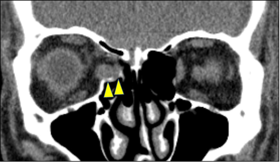
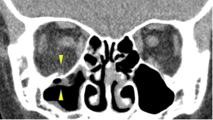
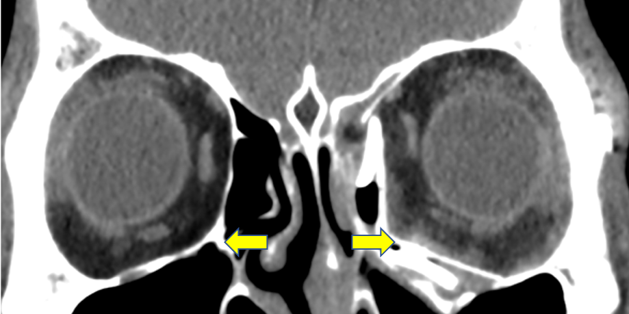
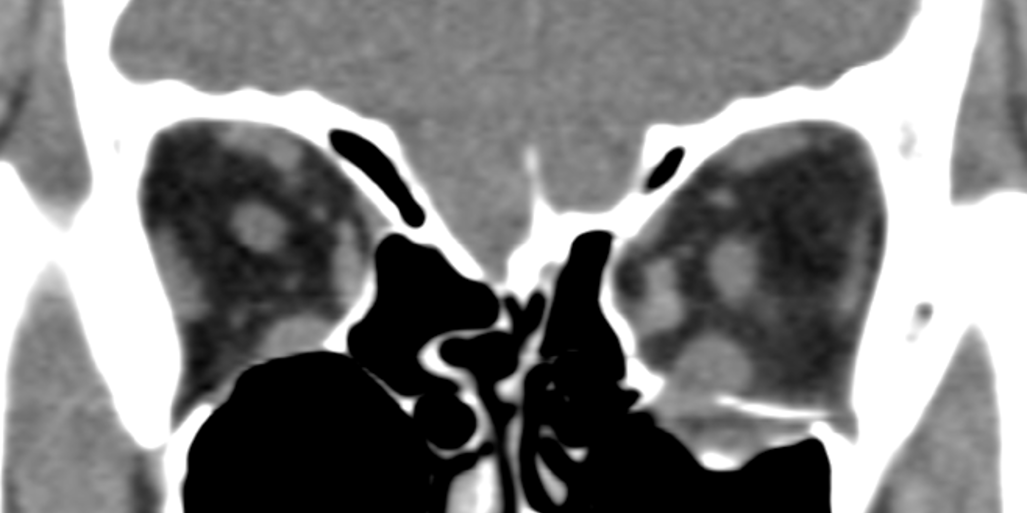
The inner wall of the orbit is deviated and protrudes into the ethmoid sinus. The medial rectus muscle appears medially deviated and swollen.
There is a hinge around the infraorbital neural canal. (Arrowhead) The broken bone is displaced into the maxillary sinus.
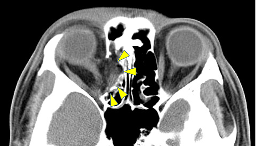
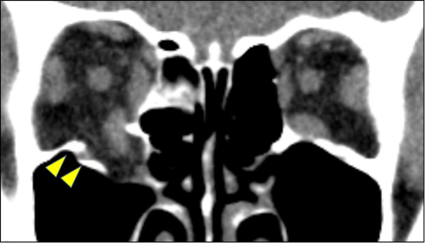
There was a fracture of the inner wall of the orbit, and the ethmoid sinus was collapsed. (Arrowhead) Orbital fat is displaced and incarcerated, and optic nerve curvature is also seen.
When an orbital fracture occurs and the orbital tissue protrudes into the space, the entire eye socket is inevitably pulled towards it, causing symptoms such as sunken eyes and a double appearance due to muscle tension.
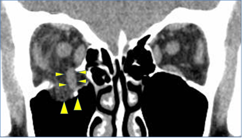
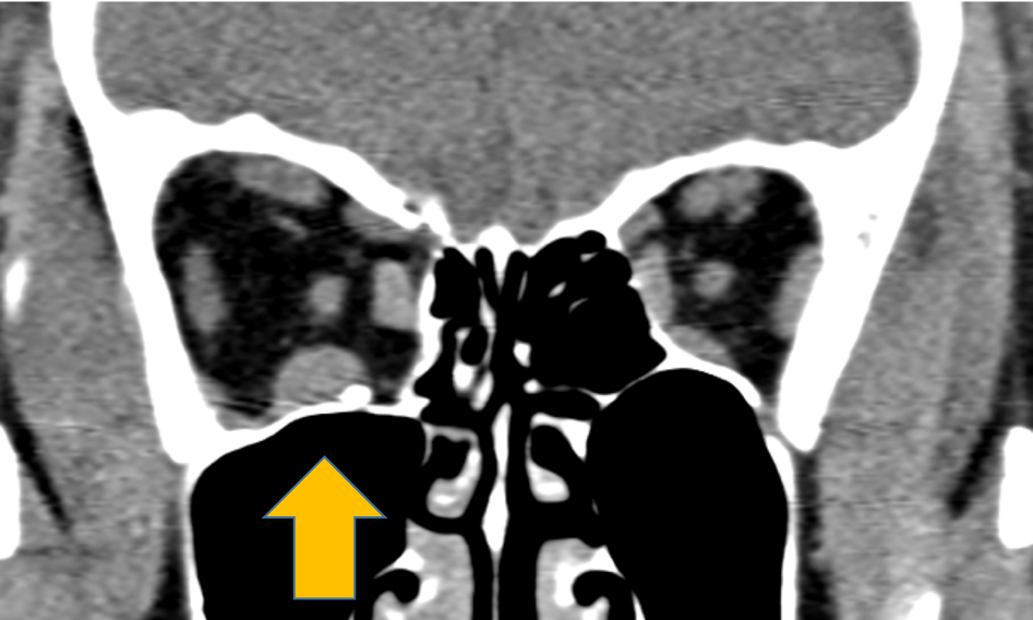
There is an open fracture, and the lower wall has fallen into the maxillary sinus. (large arrowhead)
The inferior rectus muscle is deviated and prolapsed from the orbit. (arrowhead small)
In slices further posterior to this, the inferior rectus muscle can be seen caught on the fracture edge. (arrowhead)
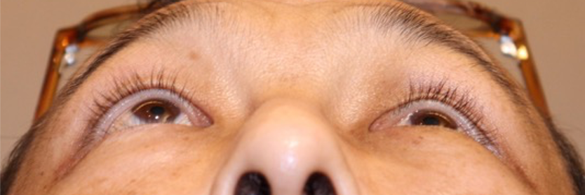
In such cases, patients complain of severe upward mobility.

This case underwent surgery at another hospital, but the orbital strut was not reconstructed, resulting in enophthalmos despite the surgery.
Of course, the best treatment is to return the eye to its pre-injury condition, so the procedure involves returning the protruded orbital tissue to its original position and reconstructing the orbital bone.
At our hospital, we heal fractures by lifting the eye from the orbital tissue side.
This is because you can return it more firmly and accurately if you pull it up from above.
<Click here for “We also cure sunken eyeballs – Orbital fracture treatment ②”>
When performing lift surgery, there are two approaches to the orbital tissue.
There are two approaches: one through a skin incision and the other through the white of the eye (conjunctiva).
At our hospital, all orbital fracture surgeries are performed starting from the white of the eye (conjunctiva).
The white of the eye (conjunctiva) is a very thin membrane that allows wounds to heal easily.
In the first place, the incision does not show up on the surface because it is located on the back of the eyelid.
<Click here for “We also cure sunken eyeballs – Orbital fracture treatment ③”>
Once you reach the orbital fat or muscle that has protruded into the space, pull it up.
The area between the orbital tissues such as fat and muscle and the bones and mucous membranes is carefully peeled away, and only the orbital tissues are returned to their original positions.
As a final step, we finish by reconstructing the bone wall, since the orbital tissue will fall back into the original space if left in place.
<Click here for “We also cure sunken eyeballs – Orbital fracture treatment ④”>
If the approach is from the white of the eye (conjunctiva), no sutures or 1-2 absorbable sutures are used, so there is no need to remove the sutures.
Apart from these, there may be cases where emergency surgery is required.
This is a condition called a closed fracture, in which the orbital bone breaks into a crack, and if muscle or fat gets caught in the crack, blood can no longer reach the bone and the tissue dies. Golden time is said to be 24 hours, so semi-emergency surgery is required.
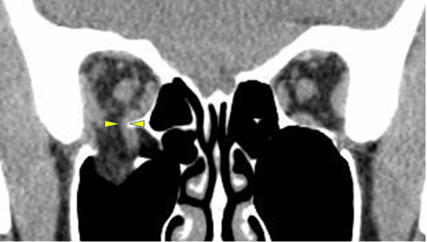
As the inferior orbital wall ruptures, the inferior rectus muscle disappears from within the orbit, a condition known as “missing rectus”.
If the extraocular muscles are involved in a closed fracture, semi-emergency surgery is indicated.
Over time, the muscles undergo necrosis, resulting in lifelong double vision.
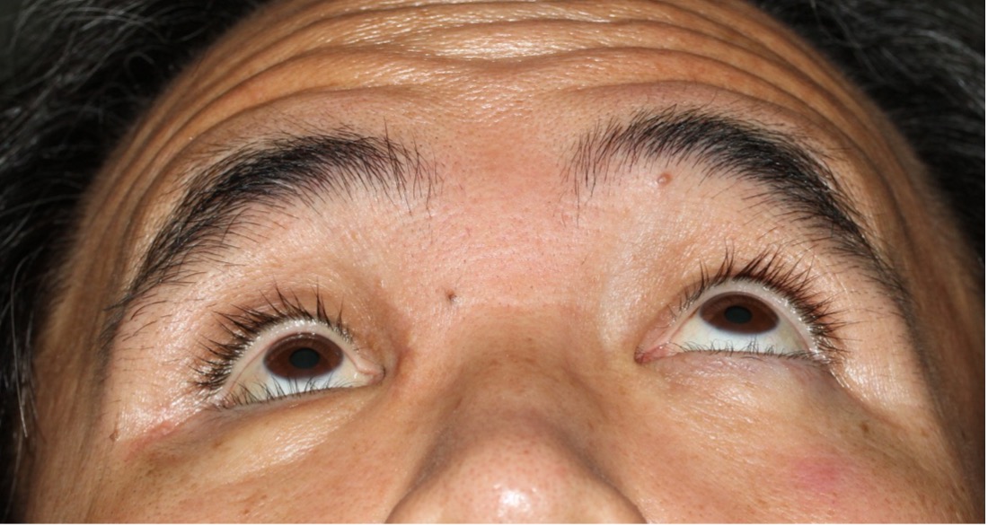
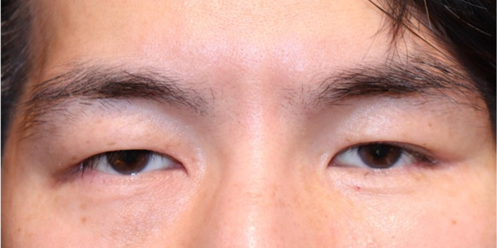
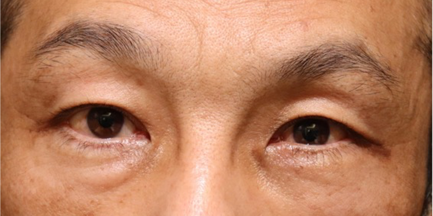
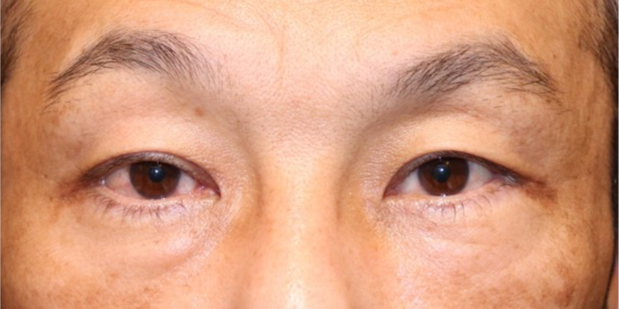
This patient have severe Enophthalmos with Orbital fracture
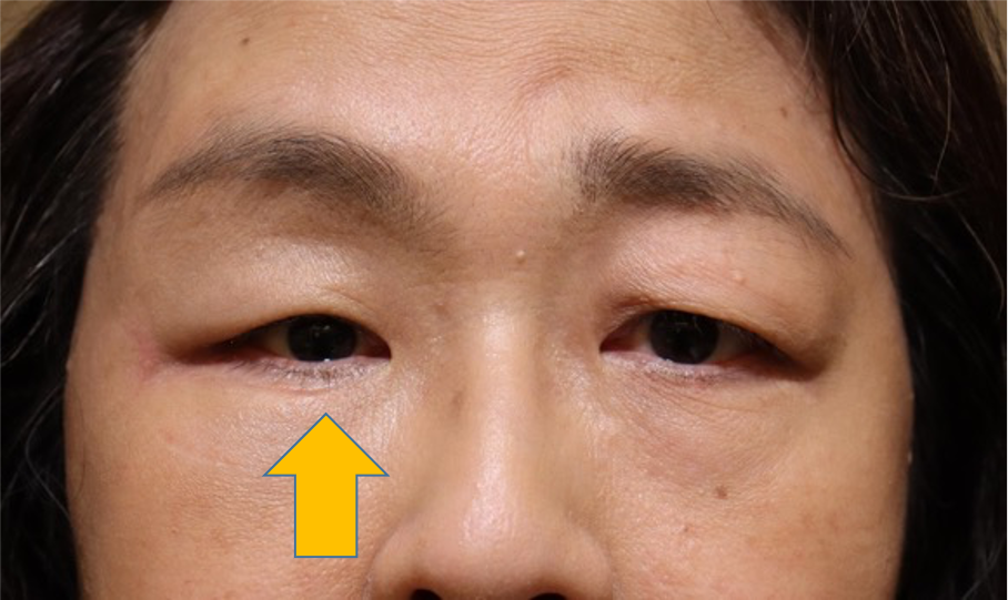
And this patient has enophthalmos with Orbital fracture too.
This patient has severe enophthalmos on the left. He was just observed until being this condition.
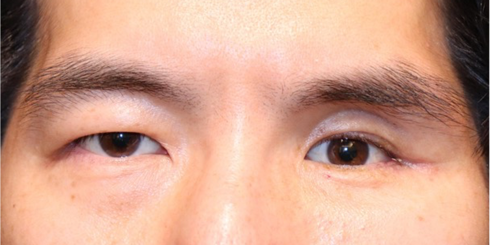
He had orbital fracture including orbital strut and medial wall and floor. Postoperatively, the position of orbital strut become symmetrical also medial wall and floor too.
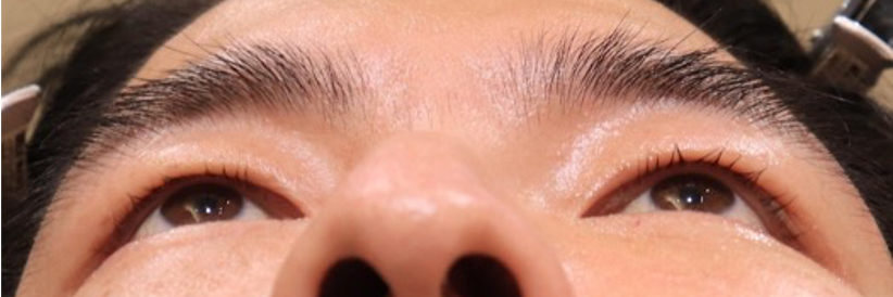
The left enophthalmos was recovered to the normal after surgery.
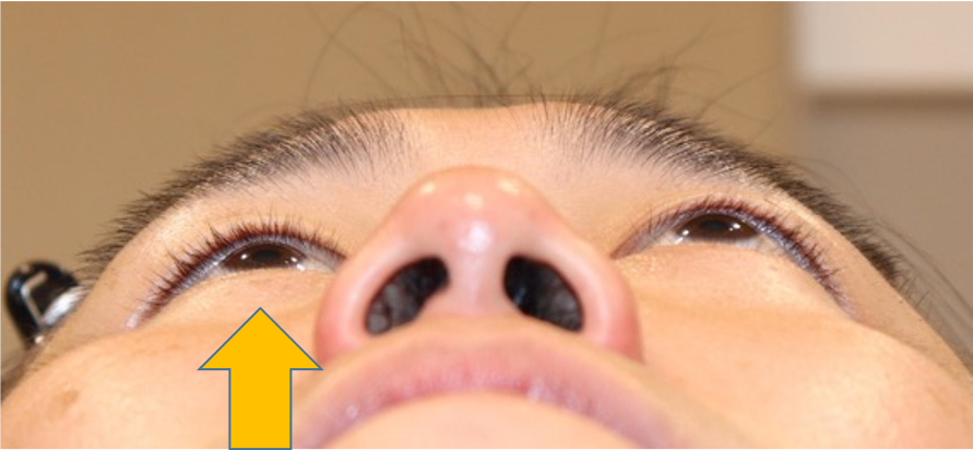
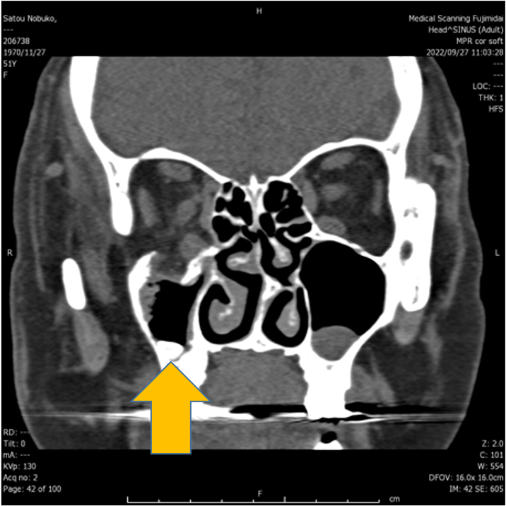
This patient has left enophthalmos on the left
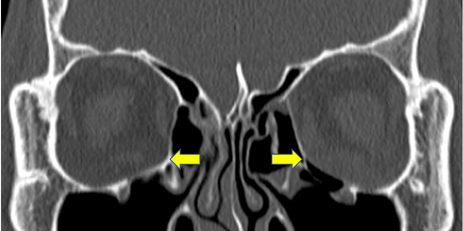
On CT scan, orbital strut including medial wall and floor was broken. So we performed repairing surgery. Postoperatively, the position of orbital strut become symmetrical.
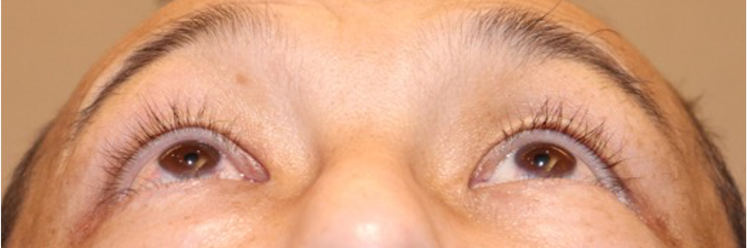
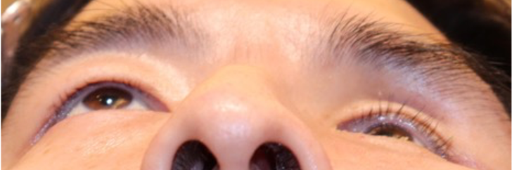
Before surgery, enophthalmos and superior sulcus exists on the left, but it disappeared after surgery.
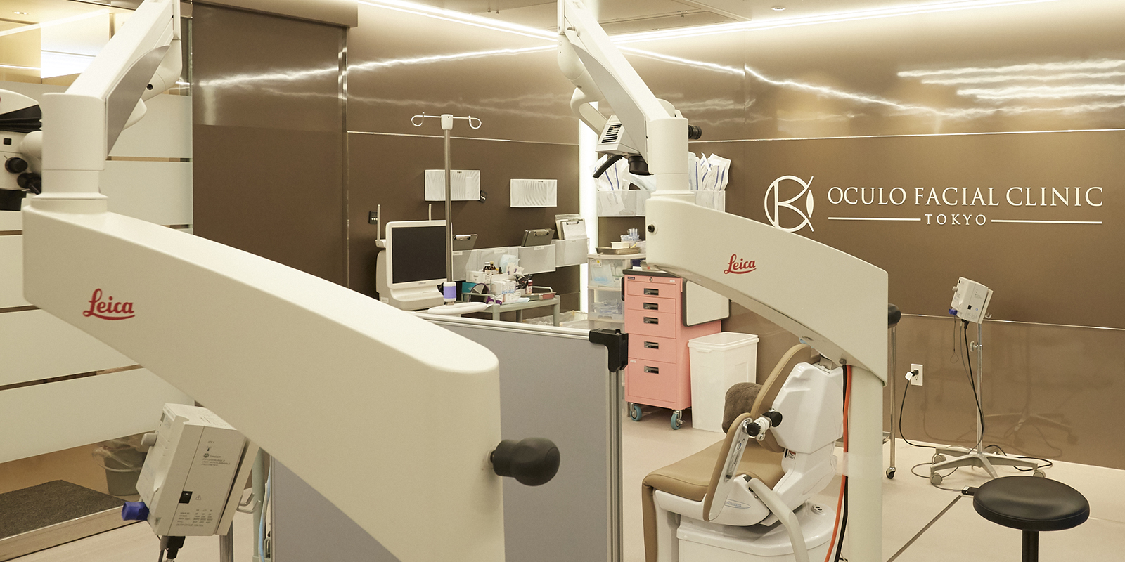
Over the past 10 years, we have performed more than 10,000 eye plastic surgeries in Japan and the United States.
I aim to provide the cutting-edge medical knowledge and experience I have gained to patients in need in Japan.
| Office Hours | MON | TUE | WEN | THU | FRI | SAT | SUN |
|---|---|---|---|---|---|---|---|
| 8:15〜17:15 | - | ◯ | ◯ | ◯ | ◯ | ◯ | - |
[Closed]Monday & Sunday ,Japanese National Holiday.
Office Hours Calendar