The major issue with Graves’ disease and thyroid eye disease is that even after receiving treatment such as drips or oral medication and entering a stable period, the appearance does not return to normal once the symptoms are resolved, unlike recovering from a common cold. Instead, the individual may end up with a completely different appearance from before. OUR GOAL IS TO RETURN THE PATIENT’S FACIAL EXPRESSION TO THE STATE IT WAS IN BEFORE THE ONSET OF THYROID EYE DISEASE.
Our goal in treatment is to restore the facial appearance before the onset of the disease.
“I want to restore my face to its original appearance as much as possible.”
Thyroid eye disease changes the appearance of people’s faces, and for some people, this can completely change their lives. I have seen many patients who become so averse to having their picture taken that they shut themselves in at home or stop going to school.
Our goal in treatment is to help patients regain their lives after they have become so affected.
Although we cannot guarantee that your face will return to its original state, we offer treatments that are not available at other medical facilities, with the aim of restoring your face to its original appearance as much as possible.
We have been fortunate to hear positive feedback from many patients. If you are worried about changes in your facial appearance, please feel free to contact us for a consultation.
“Not an Incurable Disease Anymore!? Treatment for Graves’ Ophthalmopathy The Philosophy of Oculofacial Clinic Tokyo”
Treatment of thyroid eye disease
Treatment differs between active and inactive stages.
Treatment for thyroid eye disease varies depending on the period from onset and the condition. The period immediately after onset is called the active phase, and is a state in which inflammation occurs in the muscles and fat in the orbit. Treatment for this condition is medical treatment, that is, steroid injections and infusions.
In classical treatment, steroids were also administered orally, but it has been revealed that they are less effective than injections and infusions, but have many side effects, so we do not perform this at our hospital. In addition, a new drug called teprotumumab has been released, but based on data from the United States, where it was released earlier, it is too expensive and has a low efficacy rate, so we believe that steroid treatment is more beneficial for patients.
The state in which inflammation can be calmed by administering steroids or by the passage of time is called the inactive phase, but if the eyes have not returned to their pre-onset state even after this state, surgery is indicated.
Depending on the state of the exophthalmos and the condition of the eyelids, surgery is planned with the goal of returning the patient to their original facial expression as much as possible by performing orbital decompression or eyelid surgery.
Our hospital performs orbital decompression on approximately 300 patients per year, and accepts patients from not only remote locations such as Hokkaido and Okinawa, but also from overseas.Some people experience a relapse after surgery, but the data shows that this only accounts for 2% of cases, so the risk of relapse is by no means high.
Active stage immediately after onset
Steroid injection (injection into eyelid)
Steroid drip (once a week for 4 times)
Inactive stage after steroid treatment
Orbital decompression (mainly fat removal behind the eyeball, rarely bone removal)
Eyelid surgery (to reshape the eyelids)
About steroid treatment at our hospital
Steroid treatment for active thyroid eye disease has traditionally been performed by hospitalization for three days of intravenous infusion, followed by four days of rest, repeated three times (for three weeks).
This method is effective, but it is a heavy burden for working-age patients, as it requires three weeks of hospitalization, which means being isolated from the real world for three weeks at a time.
After discharge, patients were given oral steroids, but this was a problematic treatment method due to the severe side effects and limited effectiveness. For this reason, it has recently been recommended to control the active stage of thyroid eye disease with an intravenous infusion once a week (EUGOGO).
In addition, since our hospital is a facility that specializes in orbital surgery, we use this experience to inject a steroid drug (triamcinolone) deep into the orbit.
The good thing about this injection is that it is very effective because it can be applied directly to the area of inflammation, and triamcinolone is sustained-release, so it is known to be effective for about 2-4 weeks with very few side effects (announced at the 2023 Thyroid Society).
This deep orbital injection only needs to be done every 4 weeks, so patients only need to visit the hospital once a month.
At our hospital, patients with mild to moderate thyroid eye disease receive only injections with monthly visits, and patients with severe cases receive treatment with a combination of injections and infusions with weekly visits, adopting a treatment method that places a low social burden on patients.
Treatment methods and frequency of visits for different symptoms of thyroid eye disease
Video of before and after orbital decompression surgery for thyroid eye disease Oculofacial Clinic Kashima Tomoyuki
Chief complaint
Thyroid eye disease
Treatment cost
Dr. Kashima: 1,980,000 yen
Dr. Yamana, Dr. Kikuchi, Dr. Kotaki: 880,000 yen
Other than the above *No designated surgeon (monitor required): 440,000 yen
Fat grafting to dark circles under the eyes +220,000 yen
Treatment content
Orbital decompression
Risks
Bleeding, swelling, new or worsening diplopia, vision loss, overcorrection, undercorrection
Video of before and after decompression surgery for thyroid eye disease Oculofacial Clinic Kashima Tomoyuki
Chief complaint
Thyroid eye disease
Treatment cost
Dr. Kashima: 1,980,000 yen
Dr. Yamana, Dr. Kikuchi, Dr. Kotaki: 880,000 yen
Other than the above *No designated surgeon (monitor required): 440,000 yen
Fat grafting to dark circles under the eyes +220,000 yen
Treatment content
Orbital decompression
Risks
Bleeding, swelling, new or worsening diplopia, vision loss, overcorrection, undercorrection
Surgery for Graves’ Disease and Thyroid Eye Disease
Advantage of Our Method
Only excess fat is removed
No incision of the skin
Outpatient surgery
Simultaneous surgery on both eyes
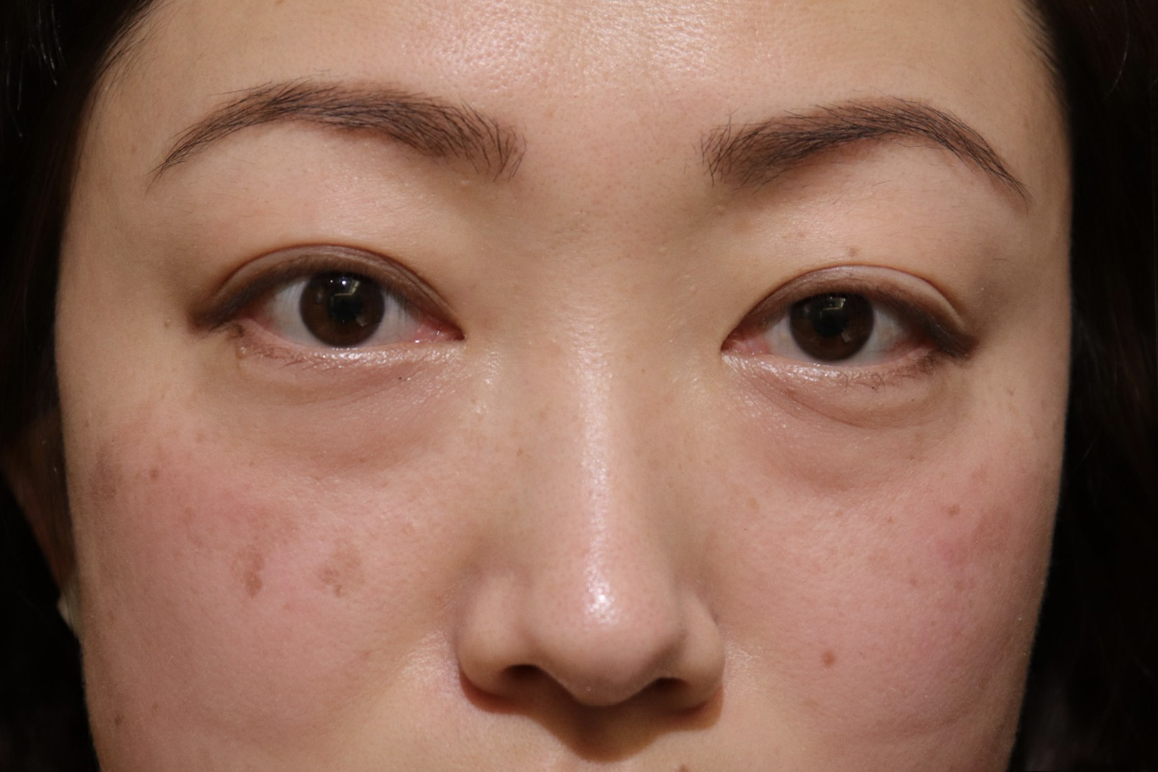
In Graves’ disease and thyroid eye disease, the fat behind the eyeball increases and the muscles that move the eyeball swell, causing the eyeball to protrude forward, resulting in significant changes to the facial appearance.
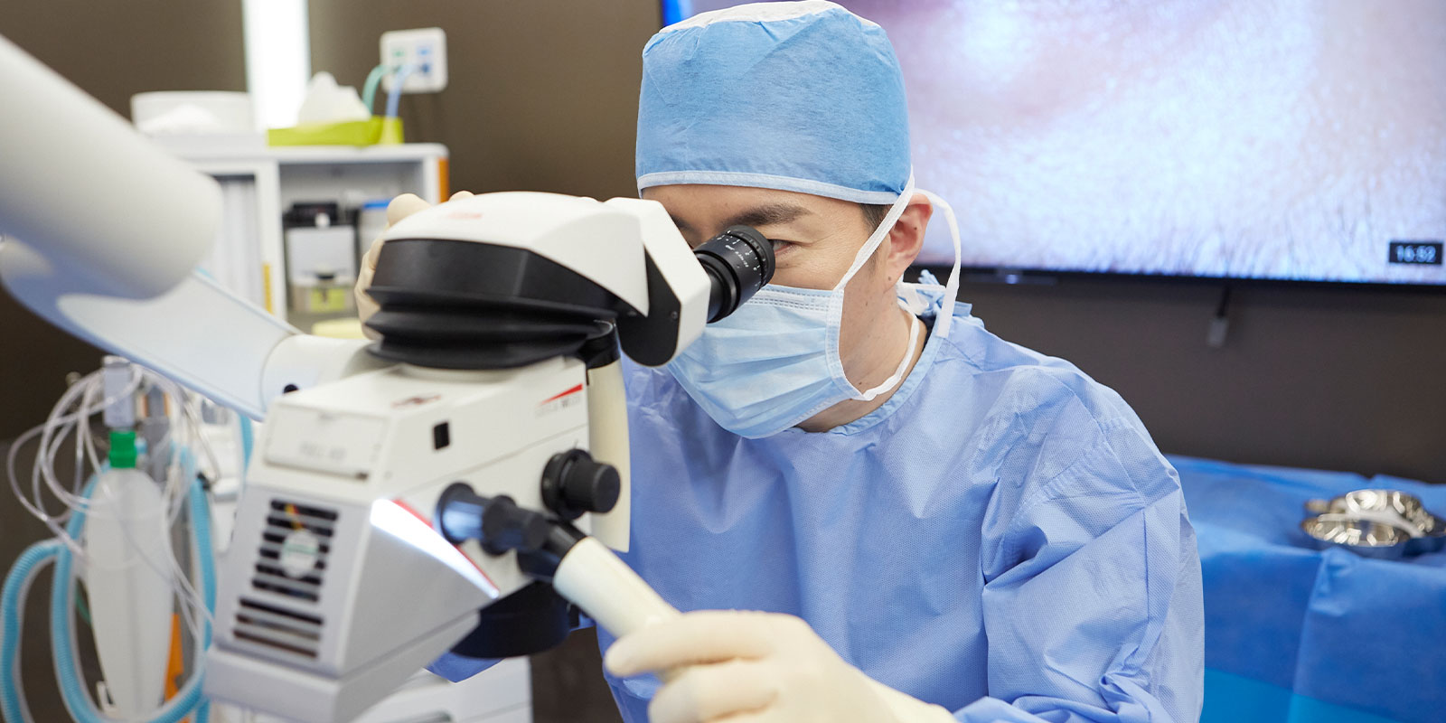
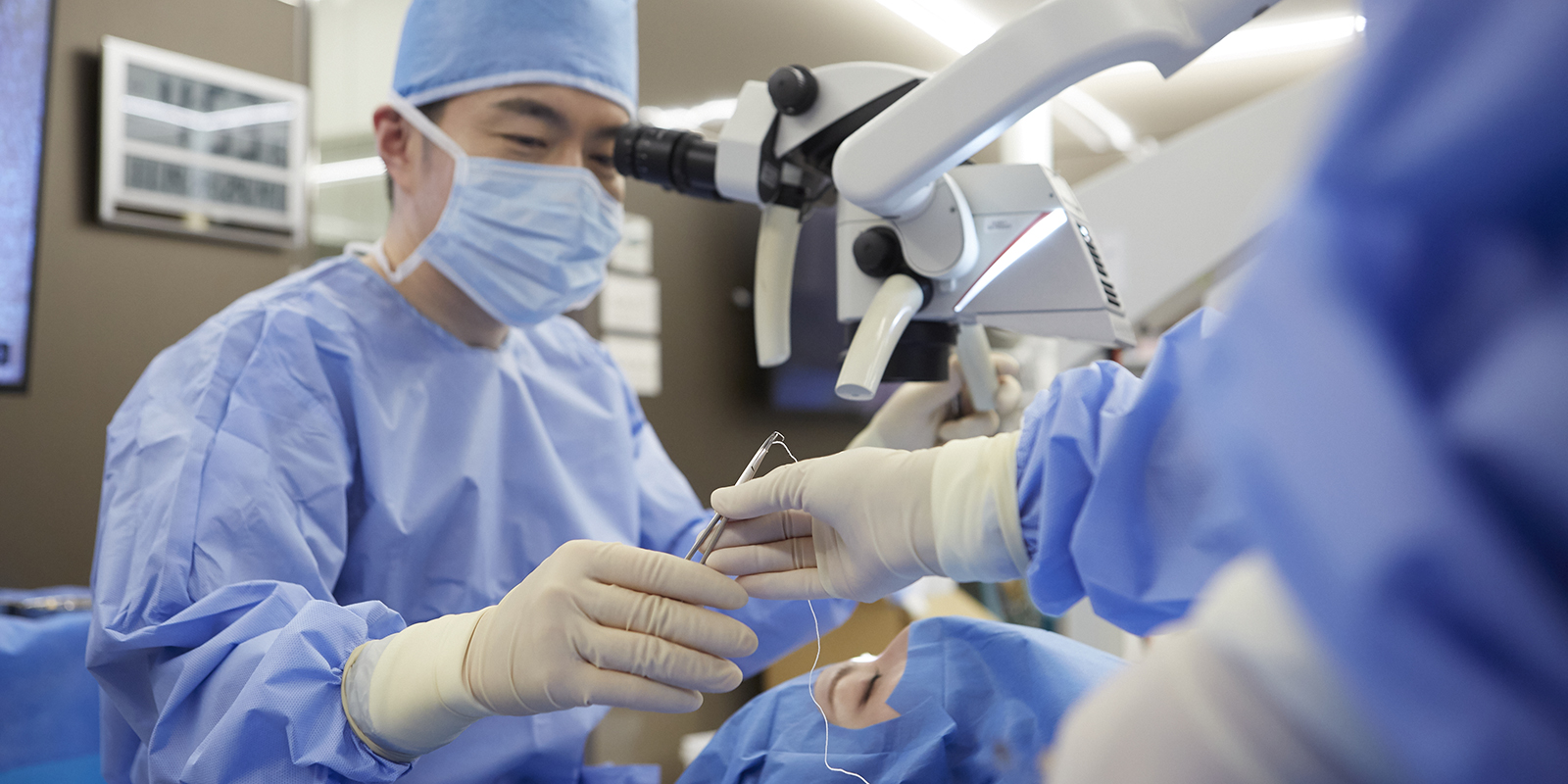
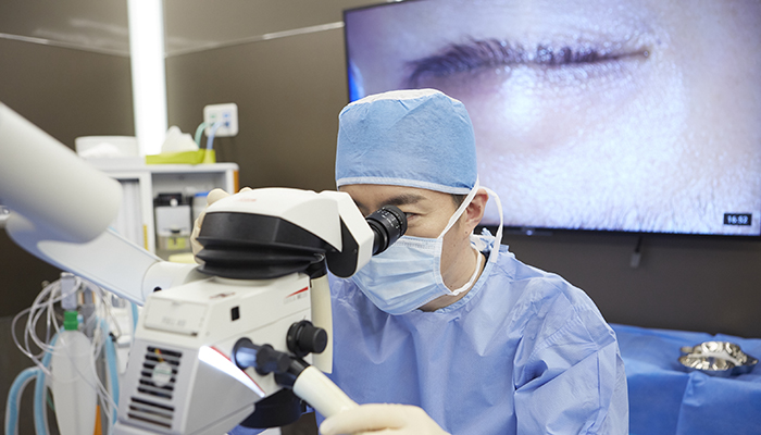
The orbital fat decompression surgery conducted at our clinic is a culmination of global expertise, incorporating cutting-edge medical knowledge acquired through studying in the United States and insights gained from renowned international experts. This surgery is meticulously performed in a Japanese style using microscopes to further minimize damage to essential tissues.
There are no medical institutions worldwide conducting this surgery.
Day surgery is possible
This surgery involves removing only the excess fat while avoiding nerves and blood vessels related to visual function, requiring highly delicate and specialized techniques. Decompression surgery involving bone removal was developed because there have been no technique to remove fat while avoiding nerves and blood vessels, but it requires significant physical burden due to bone removal, and hospitalization for 1-2 weeks is necessary due to postoperative pain and bleeding.
With our clinic’s orbital fat decompression surgery, both eyes can be operated on simultaneously under general anesthesia on an outpatient basis, allowing patients to return home on the same day. This minimizes the impact on work and home life. If you are troubled by your condition, we hope you will consider our clinic’s orbital fat decompression surgery.
both eyes/ one eye
Orbital decompression
20,000USD (Included coordinator fee)
Special price Surgery fee*1
20%OFF
*1:We offer treatment at a discounted rate for those willing to cooperate with us by allowing photography before and after the procedure, as well as video recording during the surgery, for publication on our website.
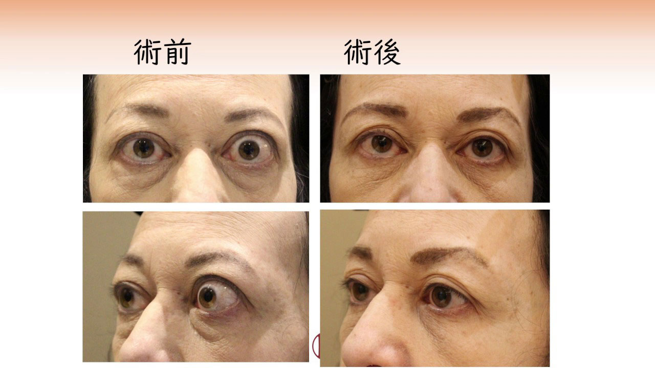
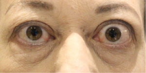
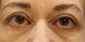
Graves’ disease and thyroid eye disease exhibit significant differences in facial appearance before and after treatment.
This woman underwent the following treatments since the onset of Graves’ ophthalmopathy:
Steroid pulse therapy
Orbital decompression surgery
Additional fat removal
Strabismus surgery (performed at another clinic)
Mainly, the above treatments were administered. When comparing before and after treatment, you can see a significant improvement, and her facial appearance looks completely different.
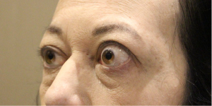
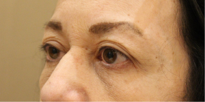
Facial profile
The difference before and after treatment is clear from surgery, and you can regain the beautiful facial features you had before onset.
It seems that Graves' ophthalmopathy was once called the "beautiful woman disease," but looking at this case, it's clear that the idea that "onset makes one beautiful" is a misconception.
Chief complaint
Graves’ ophthalmopathy
Treatment cost
Dr. Kashima: 1,980,000 yen
Dr. Yamana, Dr. Kikuchi, Dr. Kotaki: 880,000 yen
Other than the above *No designated surgeon (monitor required): 440,000 yen
Fat grafting to dark circles under the eyes +220,000 yen
Treatment content
Orbital decompression, steroid pulse, additional fat removal, strabismus surgery (at another hospital)
Risks
Bleeding, swelling, new or worsening diplopia, vision loss, overcorrection, undercorrection
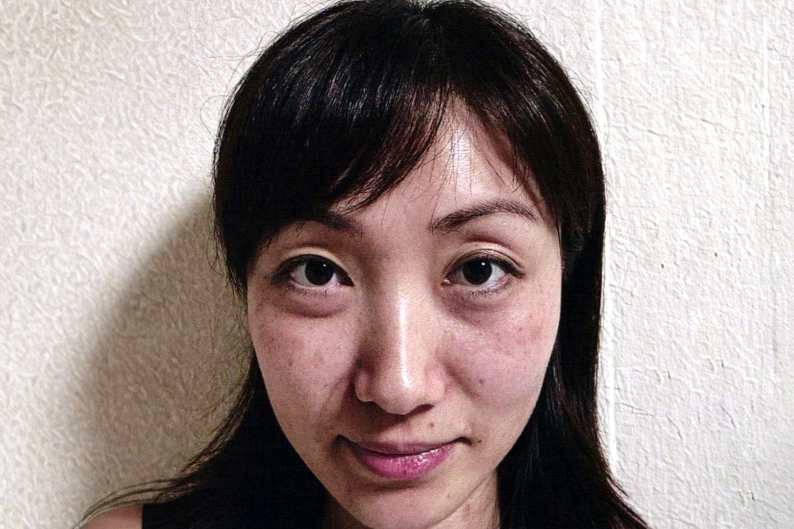
What is Graves’ disease in the first place?
A major issue with Graves’ disease and thyroid eye disease is that even if you enter a stable phase after receiving treatments like IVs and oral medications, unlike recovering from a cold, you do not return to normal but end up with a completely different appearance and physique.
The fact that these visual changes predominantly affect young women is particularly problematic. In English, this unsightly bulging of the eyes is called Disfiguring Proptosis.
There are also misunderstandings, such as “the bulging will resolve once the internal medical values stabilize,” but once it has protruded, it does not return to its original state.
Can Graves’ disease and thyroid eye disease be cured?
Many people suffer from these conditions, but there are only a few places worldwide that perform surgical treatments for Graves’ disease and thyroid eye disease.
Moreover, because the surgeries can be quite extensive within the field of ophthalmology, some facilities require several weeks of hospitalization. For this reason, there are hardly any facilities that perform surgery for relatively mild cases of eye bulging.
For such patients, we perform surgeries with a focus on cosmetic perspectives as much as possible, based on our specialized knowledge and experience.
Many celebrities have received treatments at our clinic.
Significantly improved with the latest surgical technique, “orbital decompression surgery.”
Dr. Takako Yoshida, Director of Shibuya Skin Clinic
Dr. Takako Yoshida was diagnosed with Graves’ disease at the age of 31 and subsequently developed thyroid eye disease as a complication, suffering for about 16 years.
In September 2020, she underwent surgery for thyroid eye disease at our clinic, which was successfully completed. There is a blog post about this experience, so please feel free to read it.
Shibuya Skin Clinic is a clinic for total skin care that integrates dermatology, cosmetic dermatology, and medical aesthetics. It offers a wide range of services from skin trouble treatments to home care.
If medical treatments such as corticosteroids or radiation therapy are ineffective, surgery may be necessary.
Below, I will explain the three surgical methods.
1.Orbital decompression
What is orbital decompression surgery?
Orbital decompression surgery involves the removal of fat and soft tissue or bone around the eyes to reduce the protrusion of the eyeball. This surgery removes tissue behind the eyeball, and the procedure varies depending on the part removed. The eyeball and muscles cannot be removed, so the removable parts are the increased fat and bone. The bones are divided into four directions: upper, lower, inner, and outer. Of these, the upper is close to the brain and has less decompressive effect, so it is usually not performed. The surgery typically involves the inner wall, lower wall, and outer wall. As for the fat, it is scattered around the eyeball, muscles, and nerves.
Among these, the removal of orbital fat and the outer wall of the orbit has an incidence rate of new diplopia of about 3%, but for the inner and lower walls, it ranges from 10-50%, known for a higher incidence of new diplopia. The bones of the inner and lower walls are thin, making them easy to remove, but this also means a higher risk of diplopia, which can interfere with daily activities. Furthermore, since the cheek's sensory nerve runs through the lower wall, there is a high possibility of causing numbness in the cheek.
Minimizing the burden on the body.
On the other hand, while the incidence of new diplopia is low in the removal of the lateral wall of the orbit, it is close to the brain and vital nerves and blood vessels of the eye, requiring specialized knowledge and experience to perform. Only skilled surgeons can perform this surgery.
The orbital fat decompression surgery performed at our clinic is conducted in a way that minimizes the burden on the body. We prioritize the removal of orbital fat as the first choice of surgery and avoid removing the lower and inner walls, which frequently cause diplopia, in an effort to reduce the incidence of diplopia. Also, these surgeries are primarily performed through the conjunctiva behind the eyelid or within the wrinkles of the skin, so there are almost no visible scars left. Furthermore, to reduce the number of surgeries, we perform simultaneous bilateral surgery. Local residents can go home on the same day, but for those from afar, we recommend staying at a nearby hotel after the surgery.
Surgical methods at the Oculofacial Clinic.
The primary surgical methods at the Oculofacial Clinic involve the removal of orbital fat and the shaving of the outer wall as the first choices, aiming to reduce the incidence of diplopia by avoiding the removal of the lower and inner walls, which frequently cause diplopia. Additionally, these surgeries are performed through incisions made in the conjunctiva behind the eyelid or within the skin’s wrinkles, resulting in minimal scarring. To reduce the number of surgeries, bilateral simultaneous operations are performed. Local residents may return home the same day, but for those from afar, staying at a nearby hotel post-surgery for one night only is recommended.
The day after orbital decompression! Orbital fat decompression is the best for protruding eyeballs due to Graves’ disease!
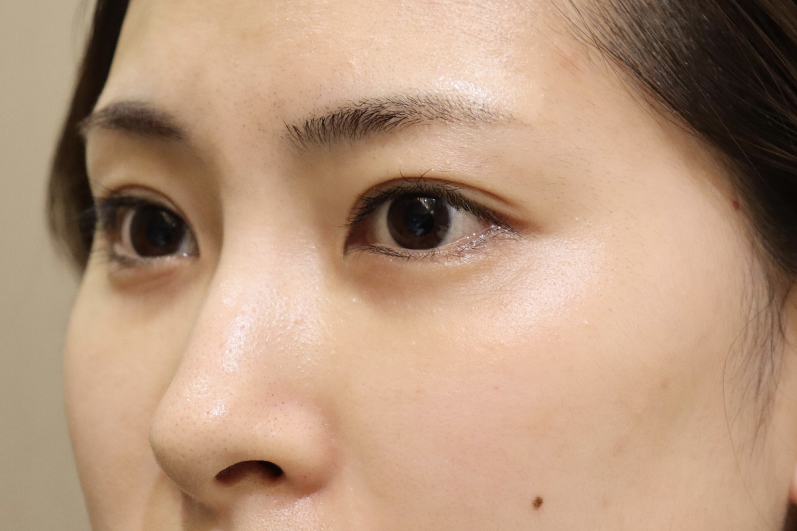
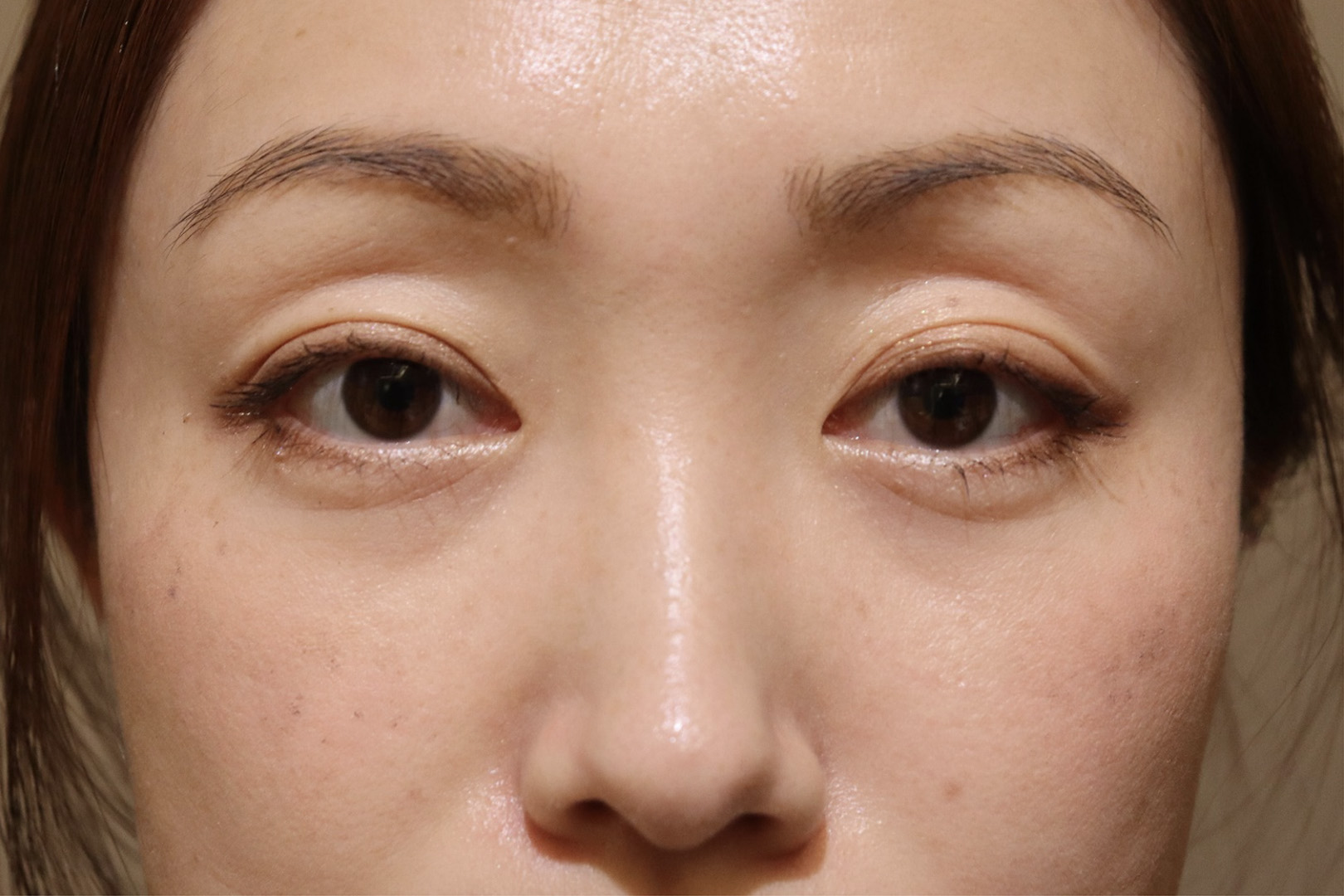
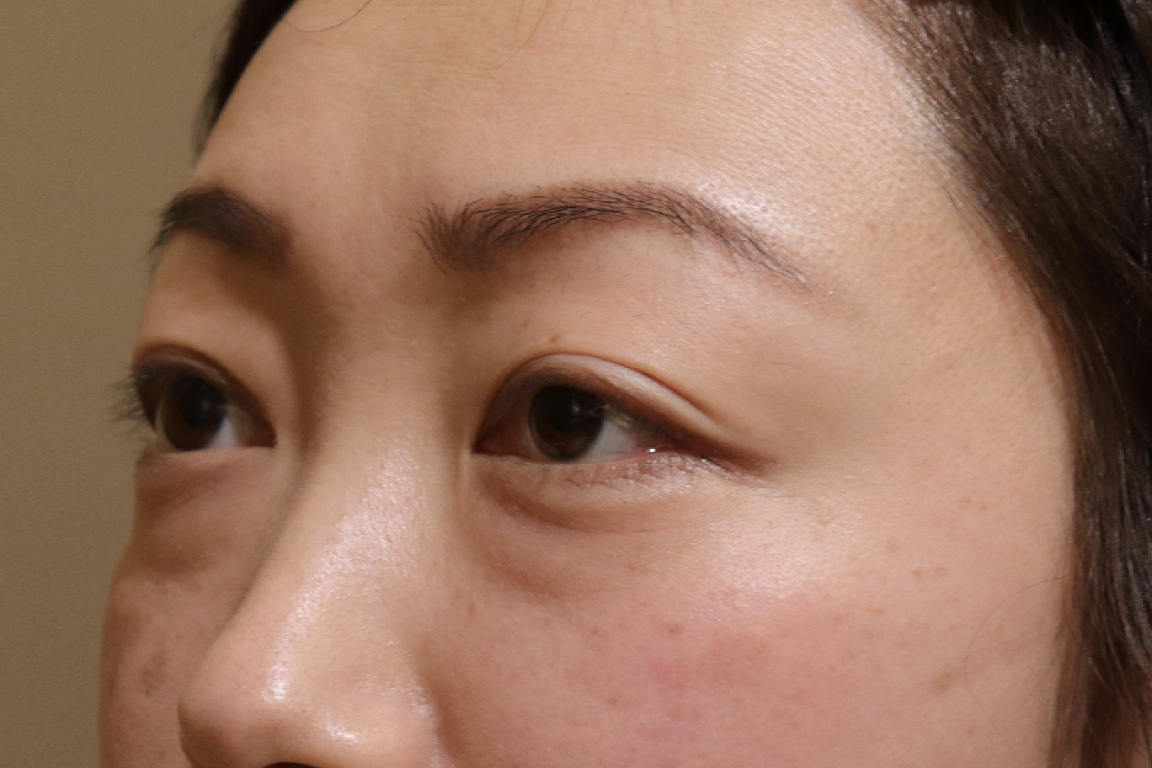
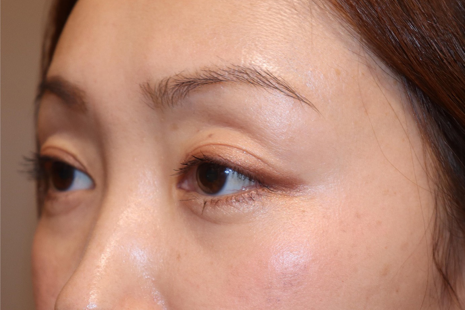
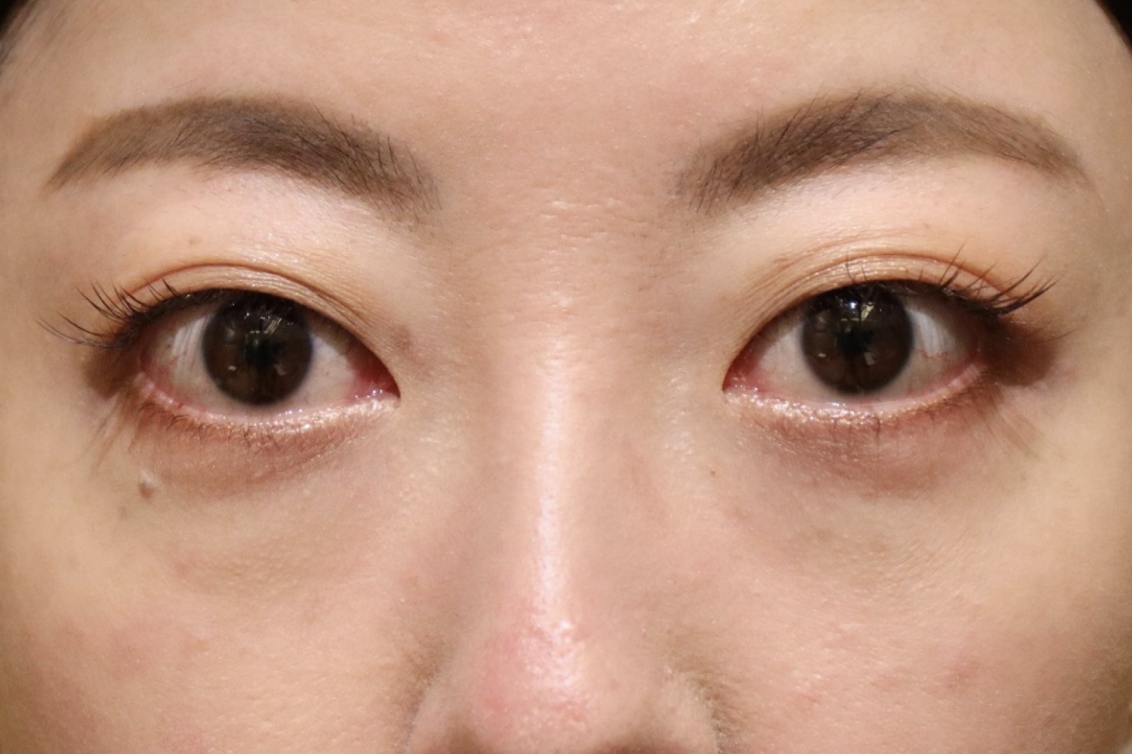
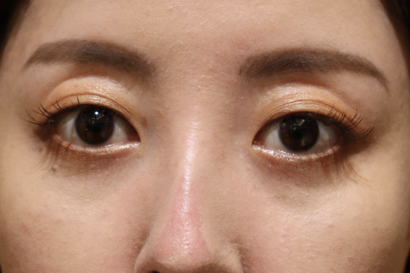
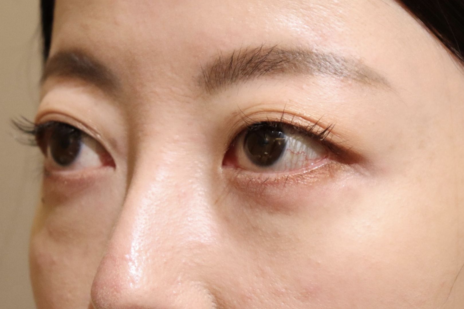
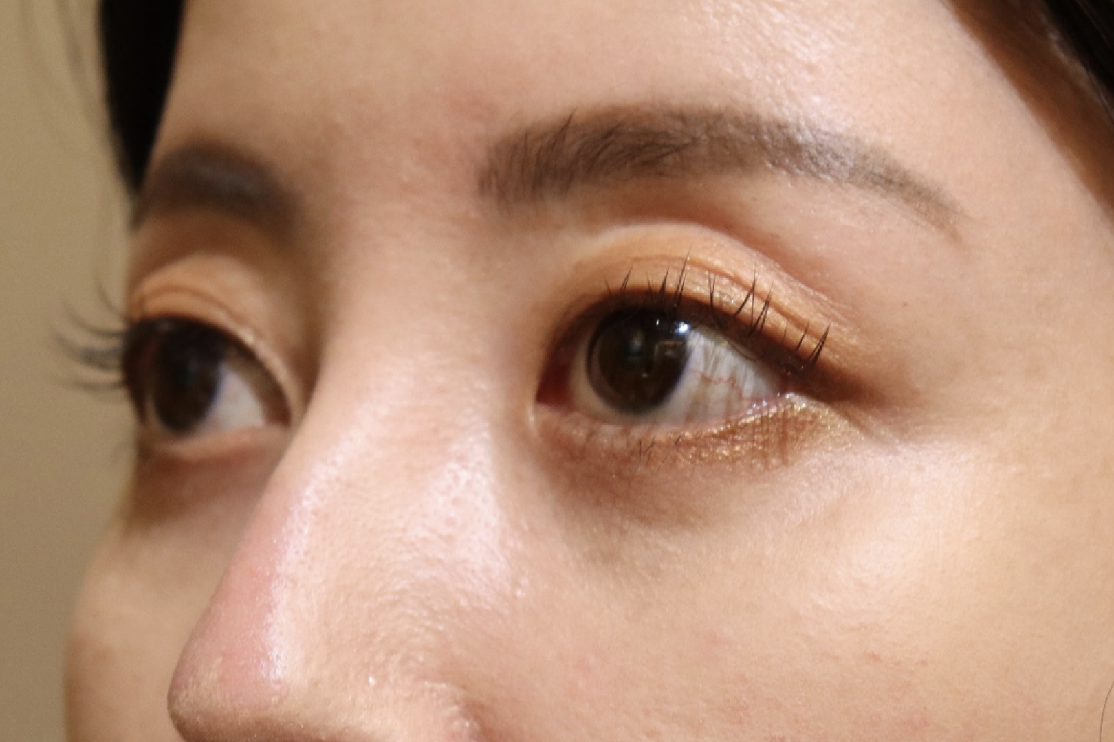
Examples of surgeries for Graves’ disease and thyroid eye disease.
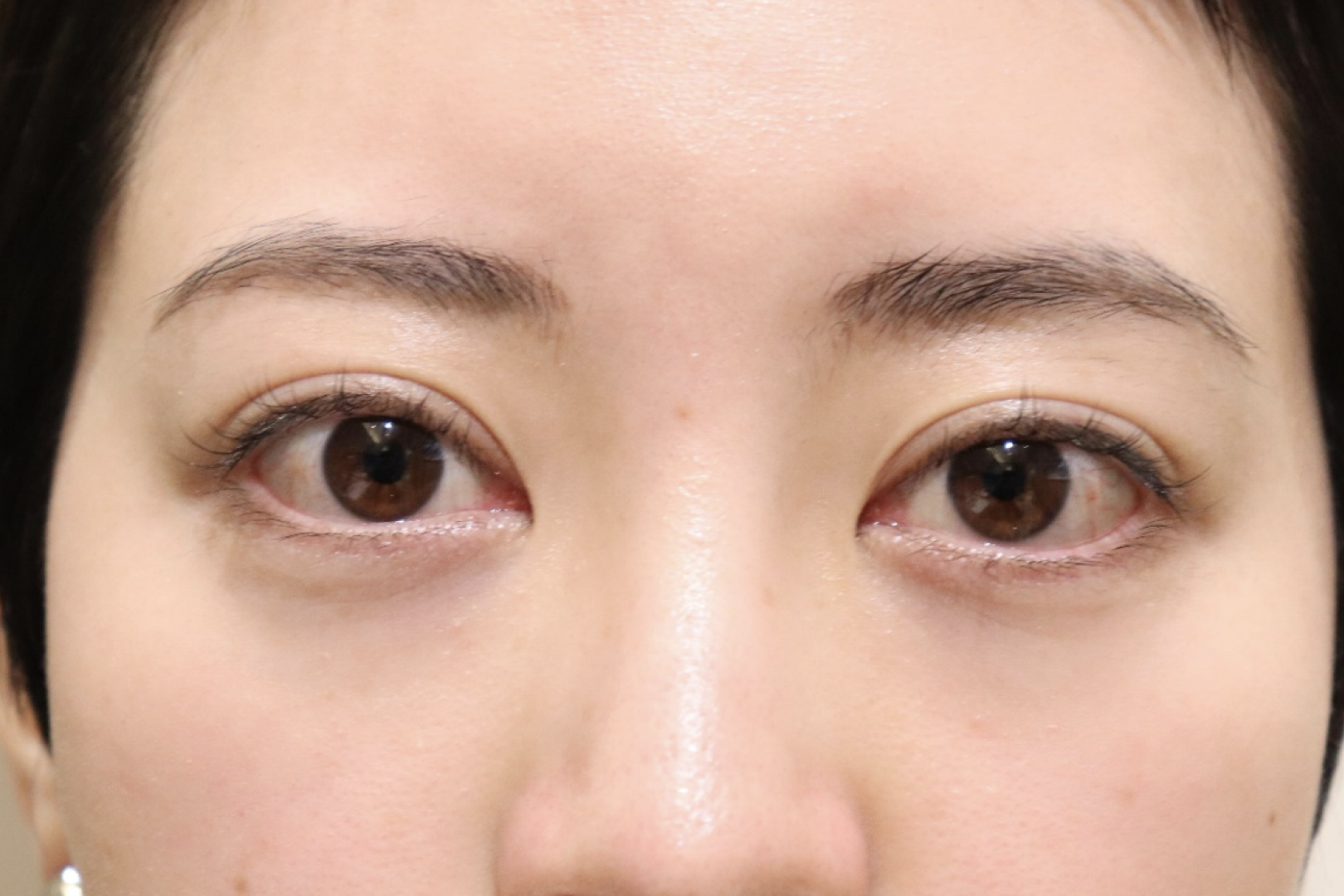
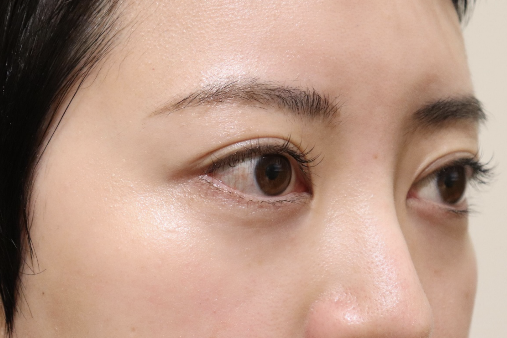
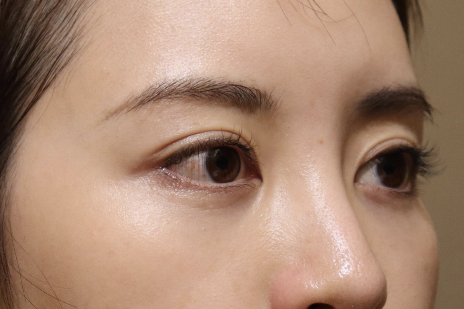
Female 20s.
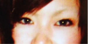
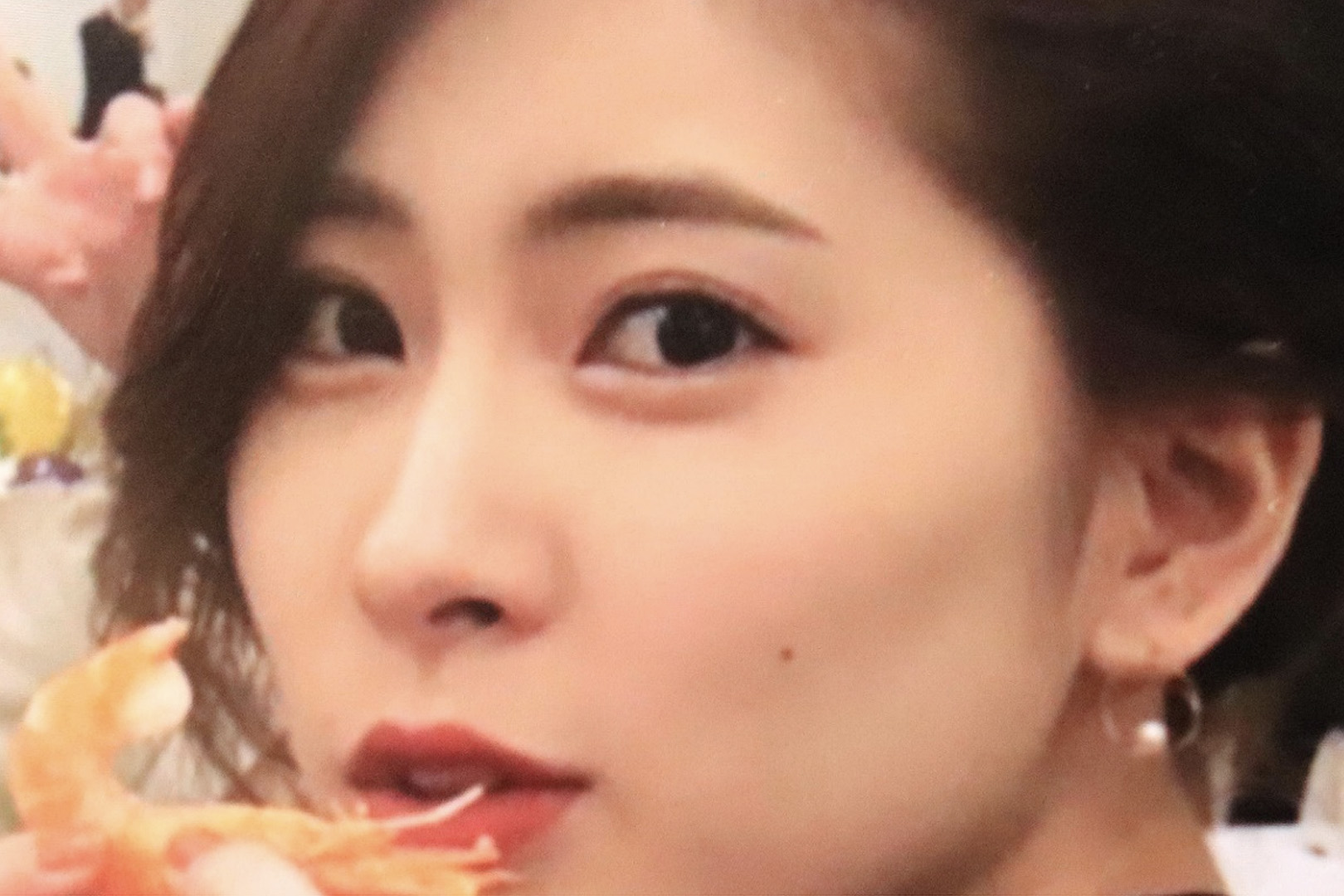
This is a photo before the onset.
The patient came to the clinic primarily complaining about changes in facial appearance and asymmetrical protrusion of the eyeballs.
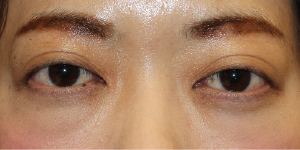
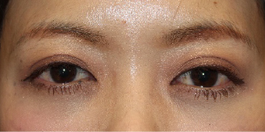
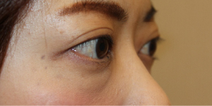
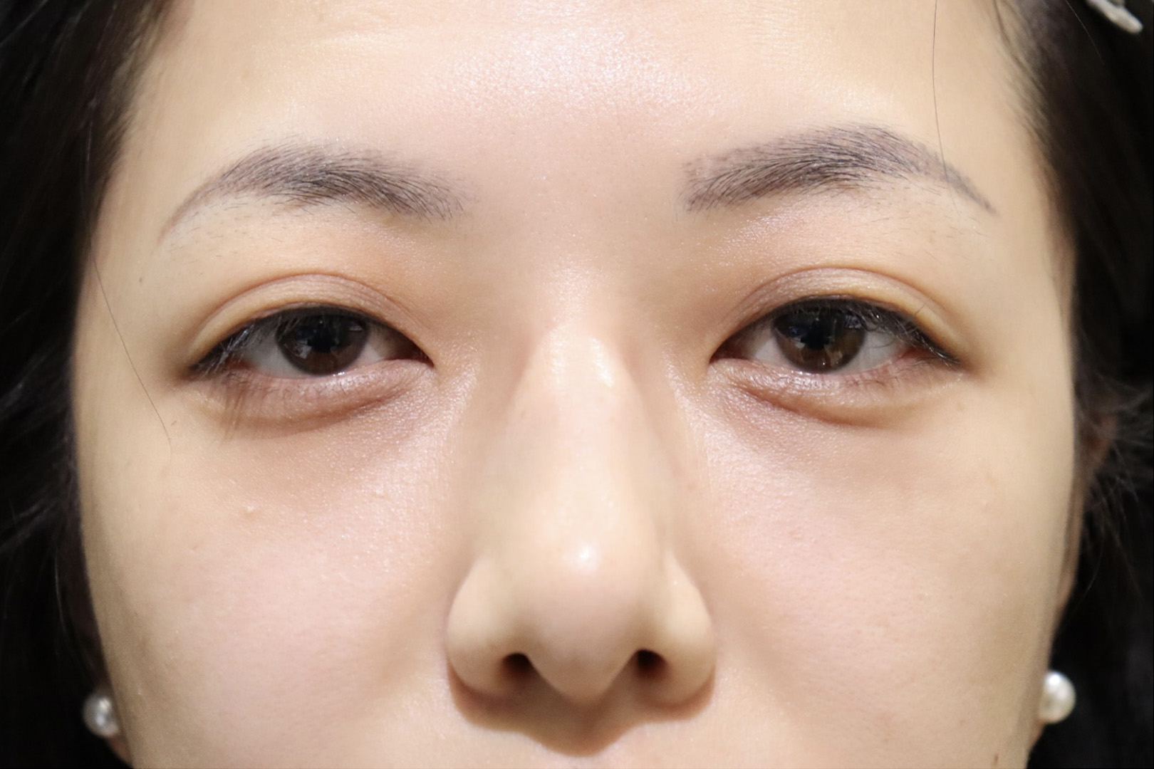
Comparison pre and post surgery
Significant improvements have been made.
From the pre-onset facial appearance, it is evident that there was a significant change to the pre-surgery appearance. The right eye is much more open, and there is a substantial increase in the exposure of the whites of the eyes both above and below. This has resulted in an asymmetrical facial appearance.
After undergoing orbital decompression surgery, there is a considerable improvement in facial appearance. Further decompression could have been performed, but since significant improvements were achieved and the patient was satisfied, the treatment was concluded.
Chief complaint
Graves’ ophthalmopathy
Treatment cost
Dr. Kashima: 1,980,000 yen
Dr. Yamana, Dr. Kikuchi, Dr. Kotaki: 880,000 yen
Other than the above *No designated surgeon (monitor required): 440,000 yen
Fat grafting to dark circles under the eyes +220,000 yen
Treatment content
Orbital decompression
Risks
Bleeding, swelling, new or worsening diplopia, vision loss, overcorrection, undercorrection
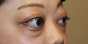
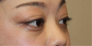
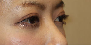
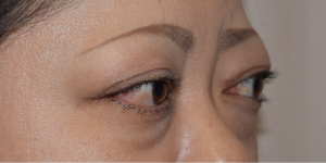
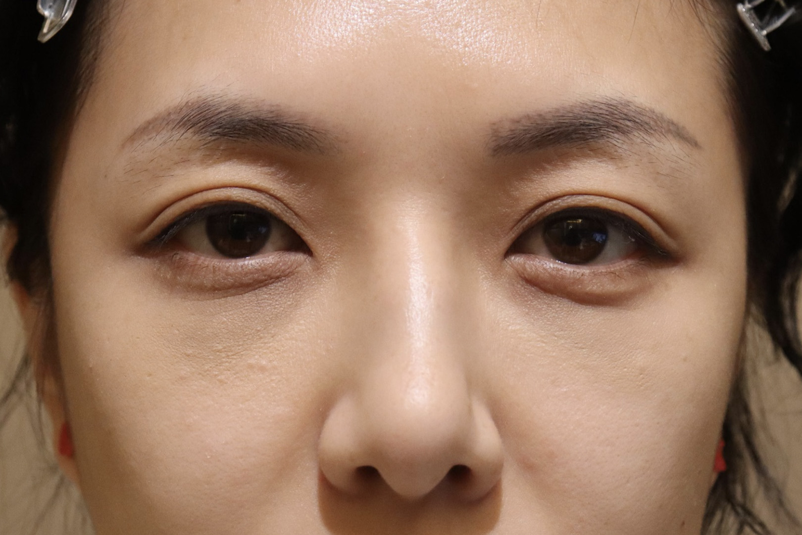
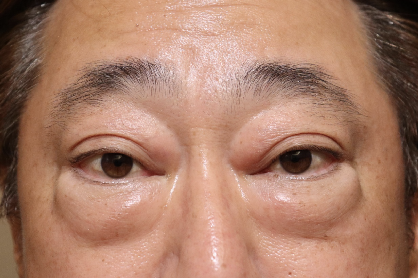
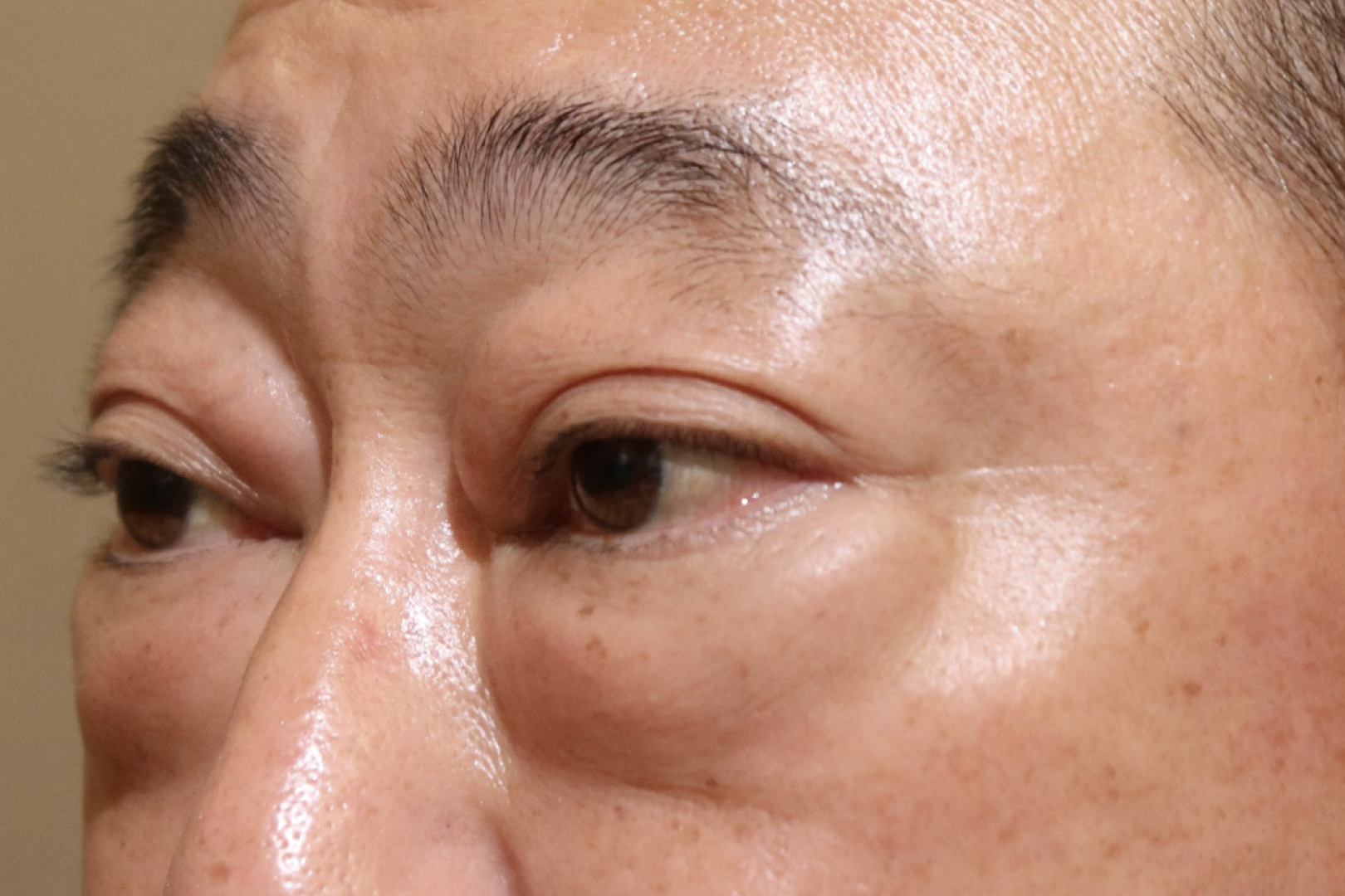
Female 40s.
The patient came to the clinic with bilateral eyeball protrusion and retraction of the upper left eyelid.
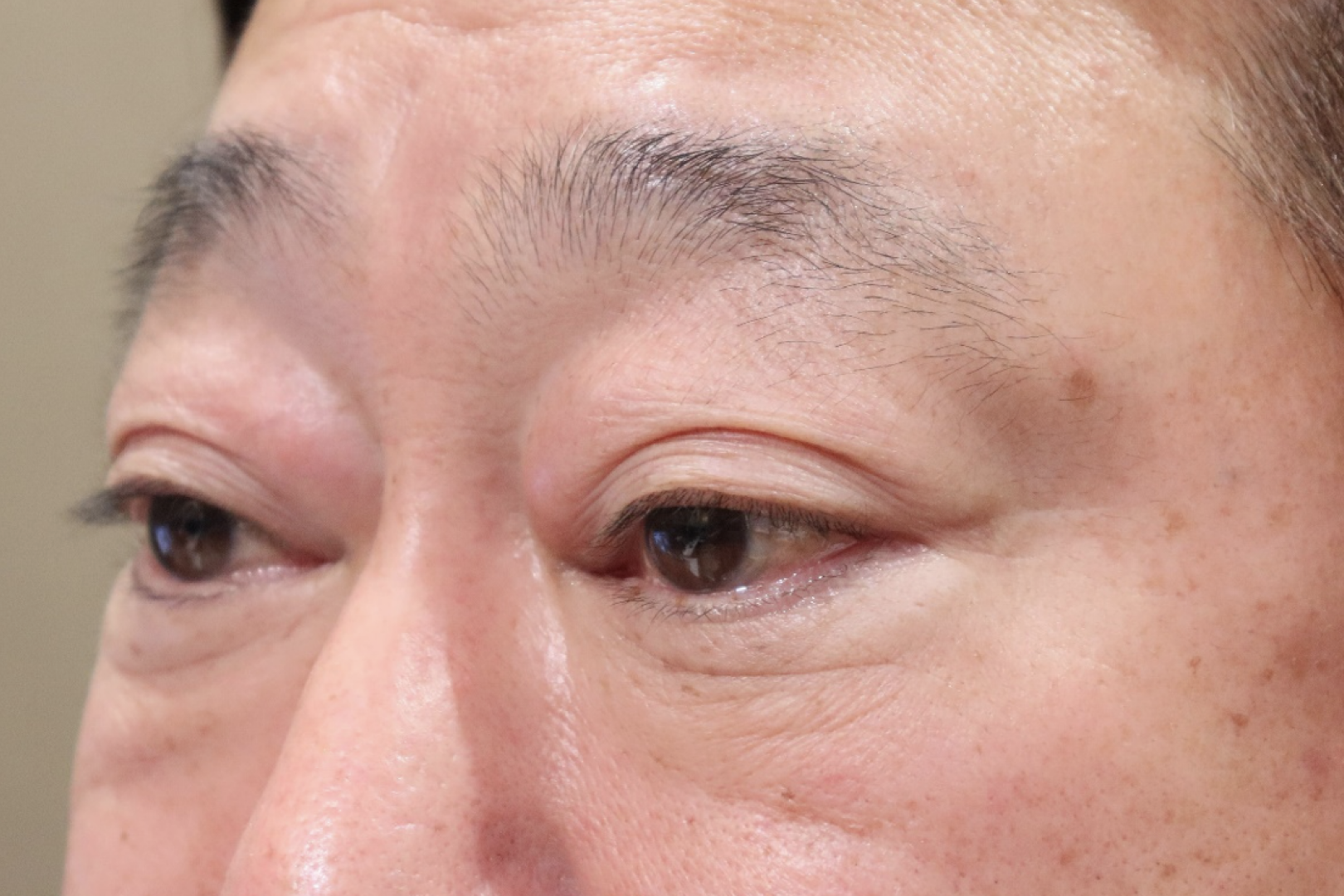
Comparison pre and post surgery
The patient’s expression has improved to a more gentle and soft appearance.
In this case, the left eye exhibited strong eyeball protrusion with visible exposure of the whites of the eyes both above and below. The asymmetry in protrusion resulted in a very pronounced asymmetry in facial appearance.
Orbital decompression surgery was performed to address this condition, followed by eyelid retraction surgery. After the surgeries, the eyeball protrusion improved, the widened appearance of the eyes was corrected, and the patient’s expression became gentler.
Chief complaint
There was bilateral exophthalmos and retraction of the left upper eyelid.
Treatment cost
Dr. Kashima: 1,980,000 yen
Dr. Yamana, Dr. Kikuchi, Dr. Kotaki: 880,000 yen
Other than the above *No designated surgeon (monitor required): 440,000 yen
Fat grafting to dark circles under the eyes +220,000 yen
Treatment content
Orbital decompression
Risks
Bleeding, swelling, new or worsening diplopia, vision loss, overcorrection, undercorrection
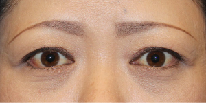
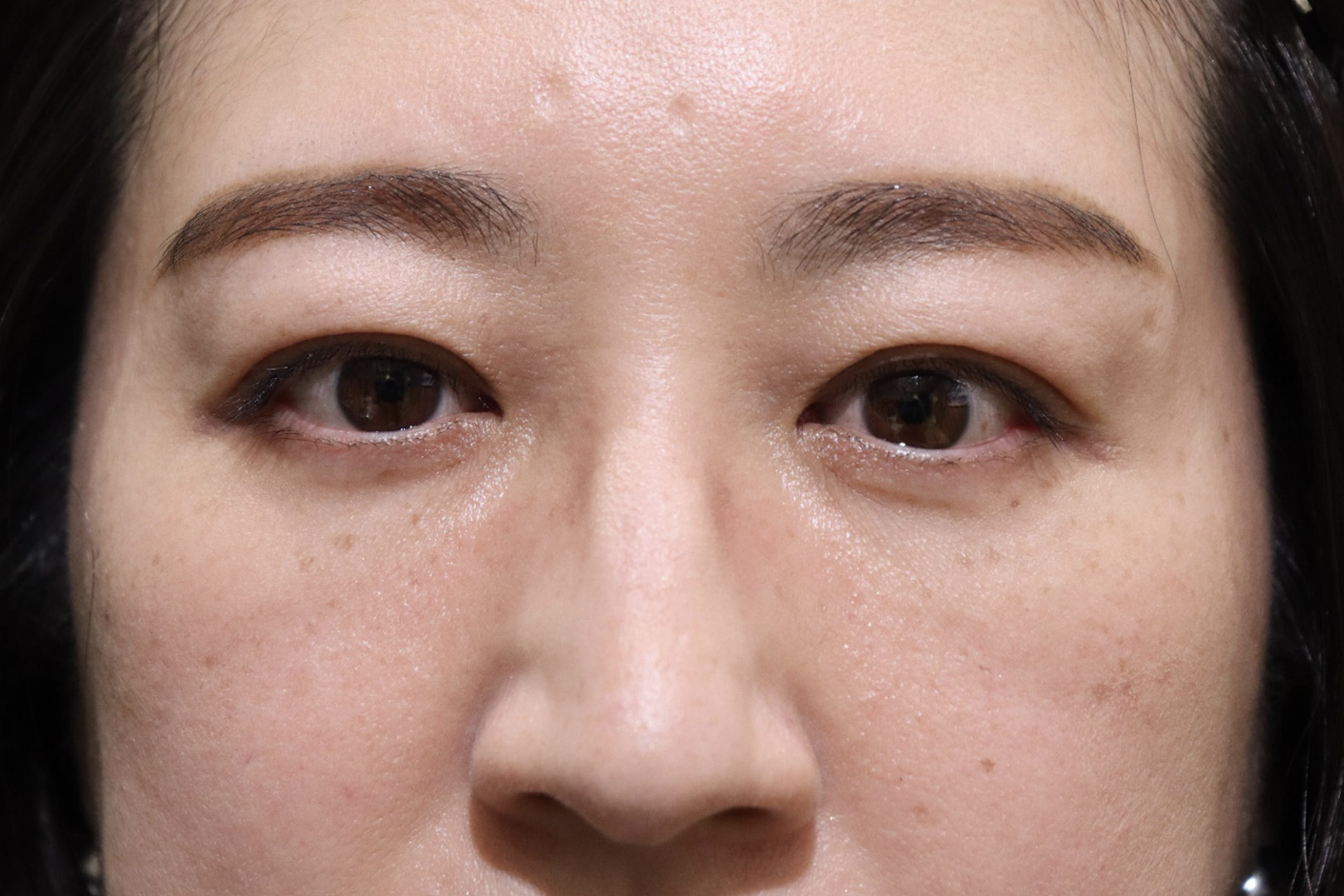
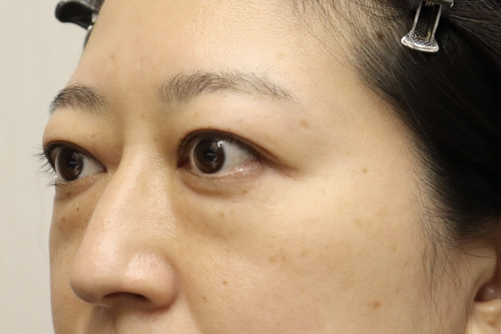
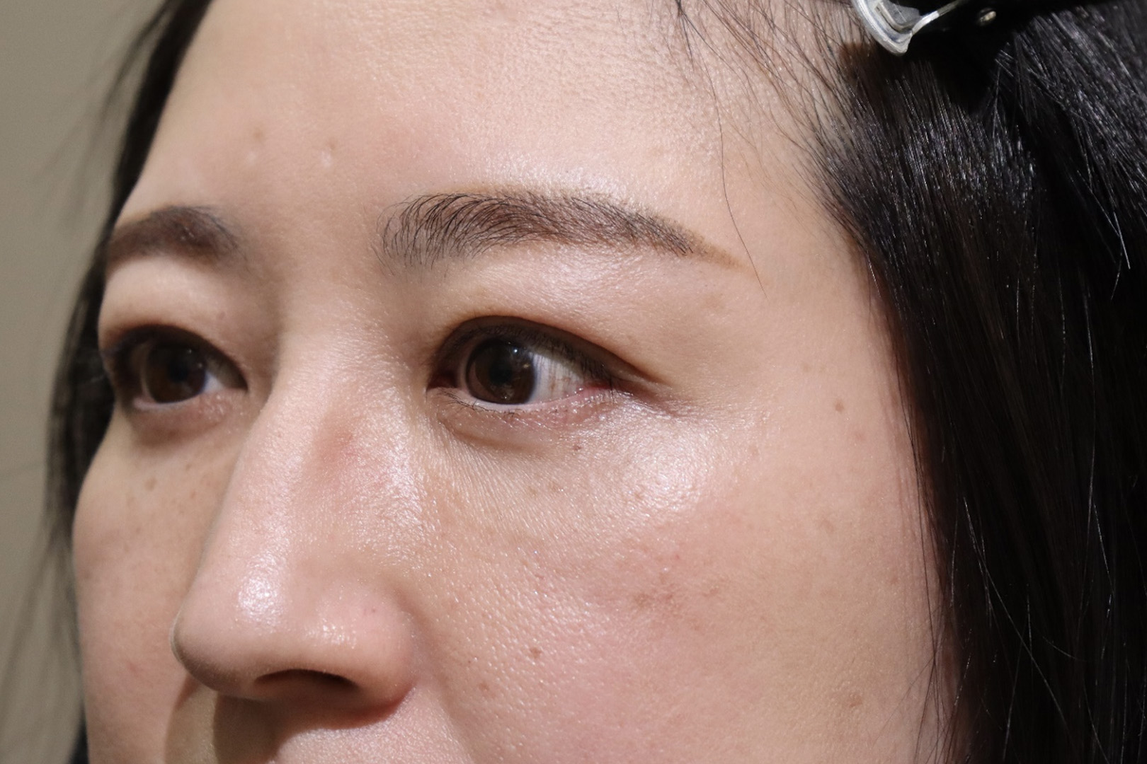
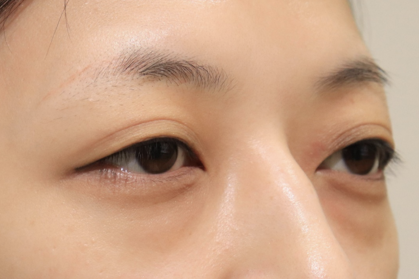
Female 30s.
This is a photo before the onset.
The patient came to the clinic primarily complaining about changes in facial appearance and asymmetrical protrusion of the eyeballs.
Comparison pre and post surgery
The swelling of the eyelids has disappeared.
It is evident that the orbital decompression surgery has resulted in a more beautiful facial appearance. Physically, the puffiness of the upper eyelids has disappeared, and the double eyelid lines have become more defined.
Chief complaint
Graves’ ophthalmopathy
Treatment cost
Dr. Kashima: 1,980,000 yen
Dr. Yamana, Dr. Kikuchi, Dr. Kotaki: 880,000 yen
Other than the above *No designated surgeon (monitor required): 440,000 yen
Fat grafting to dark circles under the eyes +220,000 yen
Treatment content
Orbital decompression
Risks
Bleeding, swelling, new or worsening diplopia, vision loss, overcorrection, undercorrection
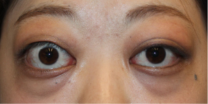
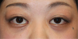
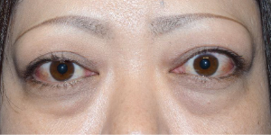
Female 40s.
Orbital decompression surgery was performed.
Comparison pre and post surgery
The swelling has gone down.
It can be observed that the swelling of the upper and lower eyelids, which was present preoperatively due to Graves’ ophthalmopathy, has disappeared postoperatively. Reduction in proptosis also improves dry eye symptoms such as redness, grittiness, and tearing.
Chief complaint
Graves’ ophthalmopathy
Treatment cost
Dr. Kashima: 1,980,000 yen
Dr. Yamana, Dr. Kikuchi, Dr. Kotaki: 880,000 yen
Other than the above *No designated surgeon (monitor required): 440,000 yen
Fat grafting to dark circles under the eyes +220,000 yen
Treatment content
Orbital decompression
Risks
Bleeding, swelling, new or worsening diplopia, vision loss, overcorrection, undercorrection
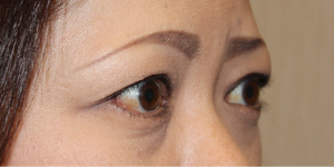
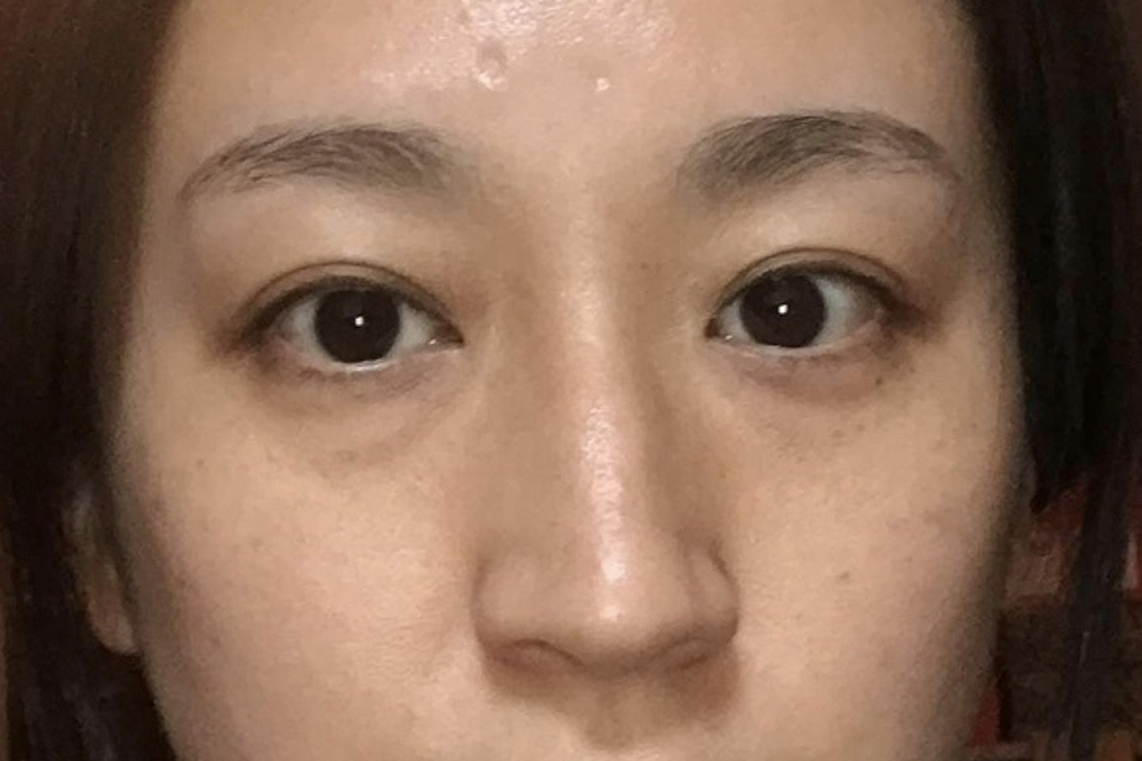
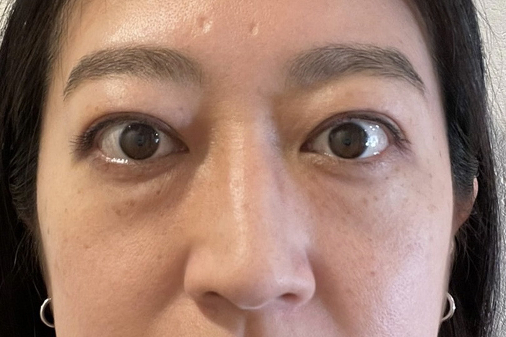
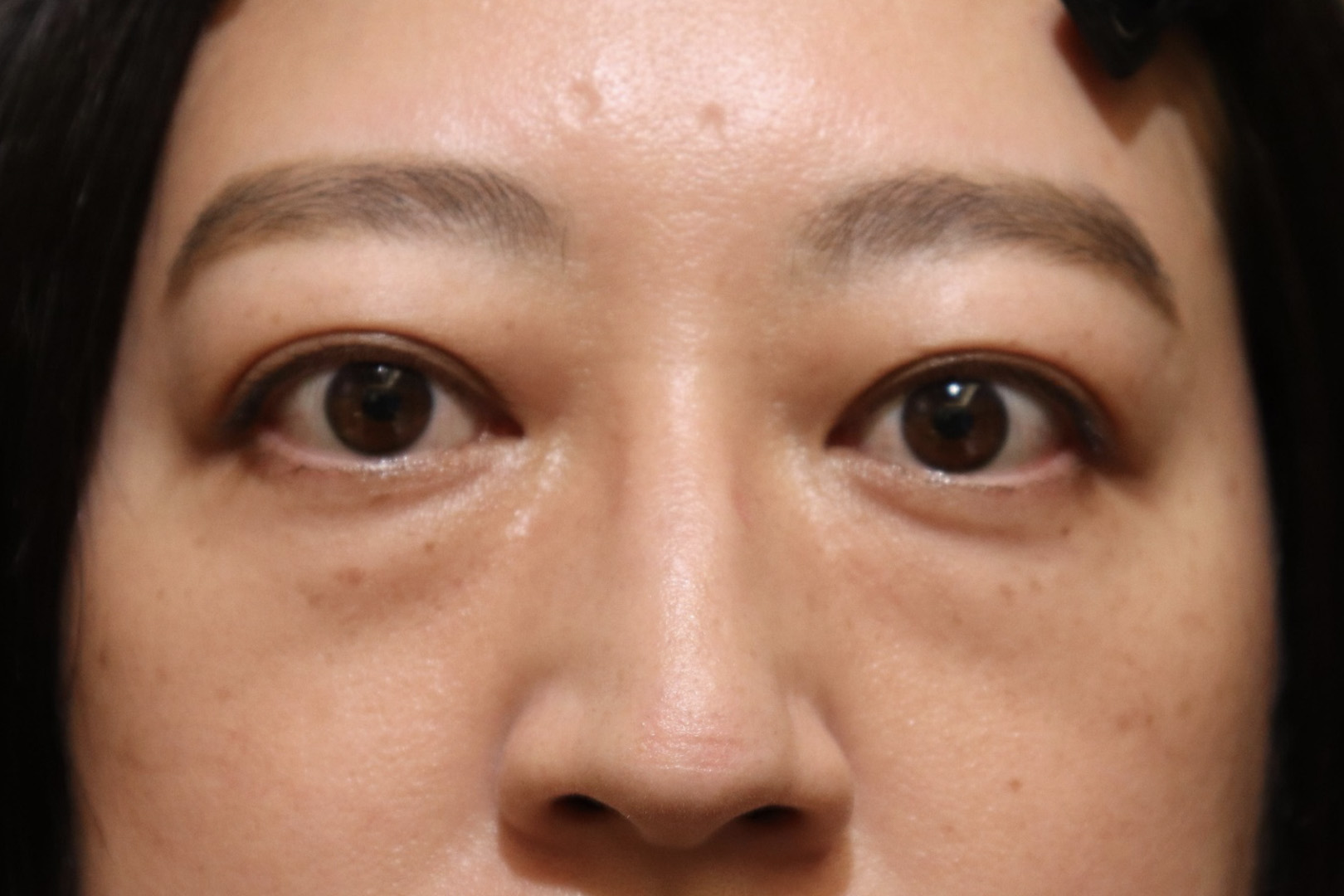
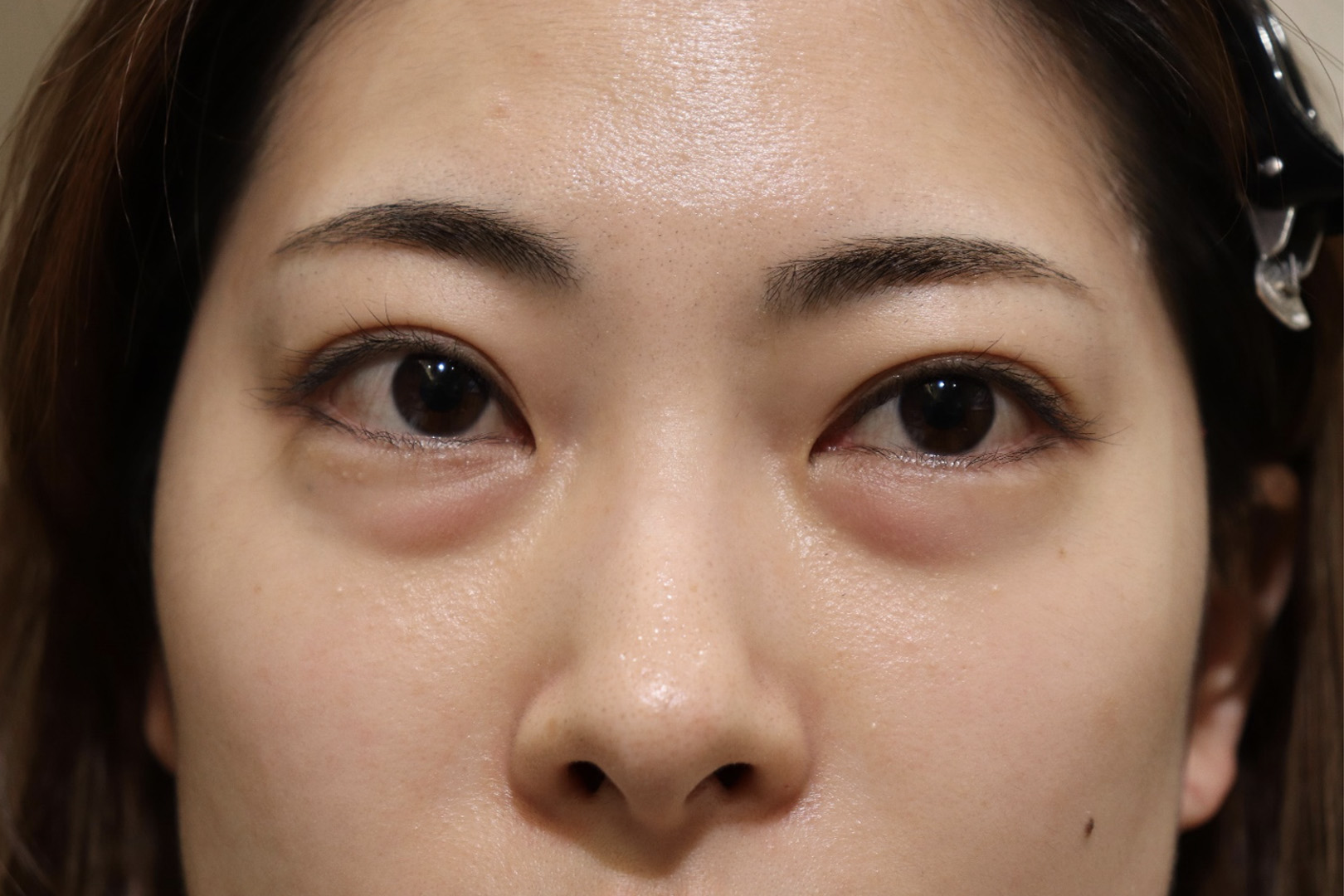
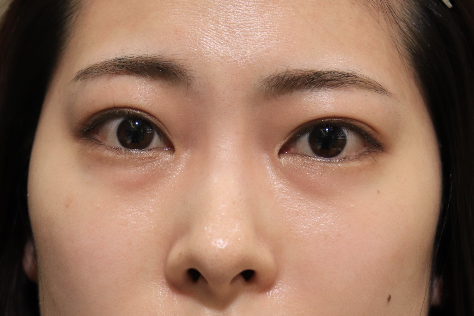
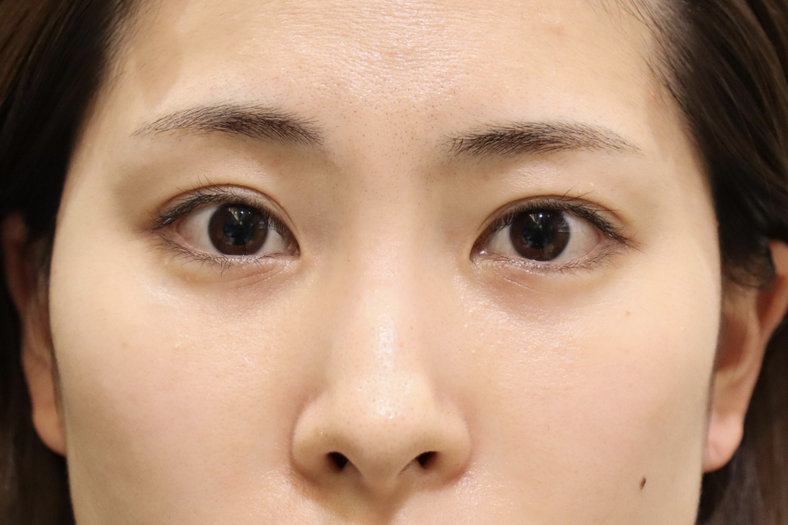
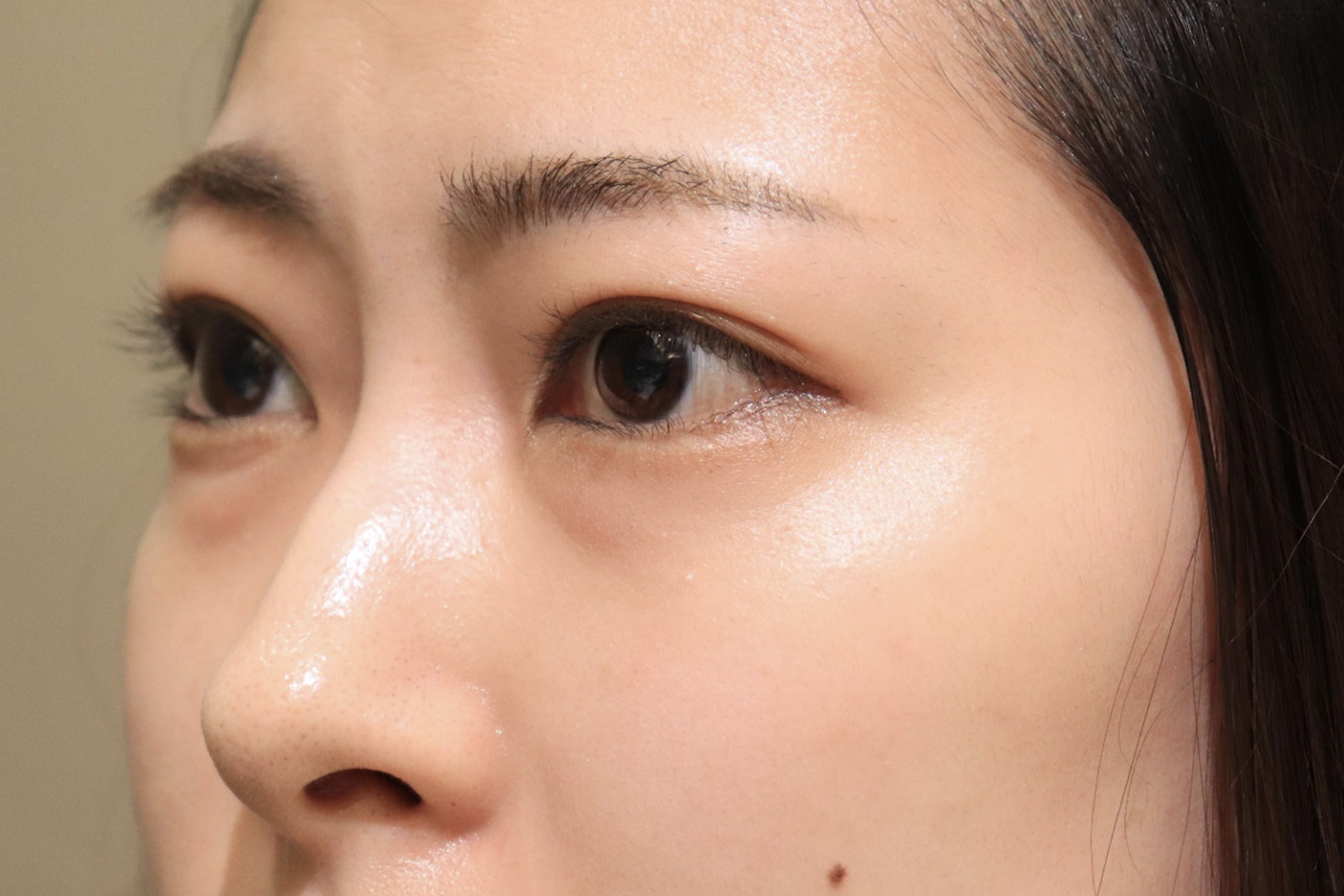
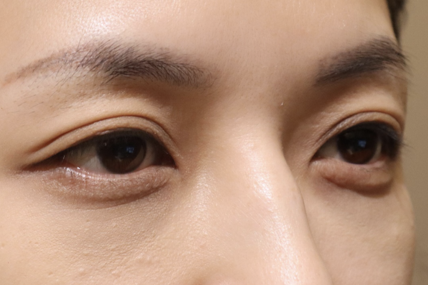
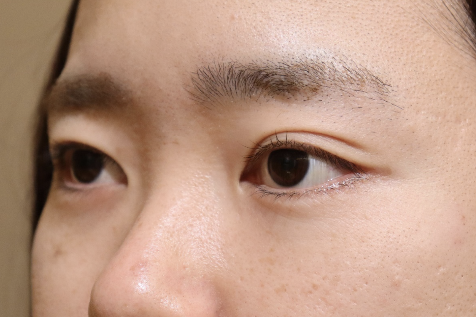
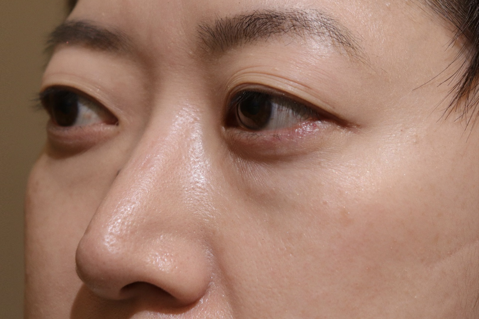
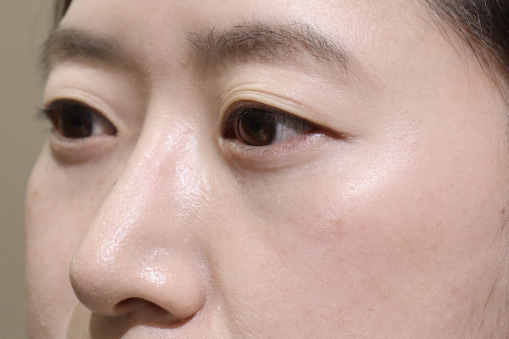
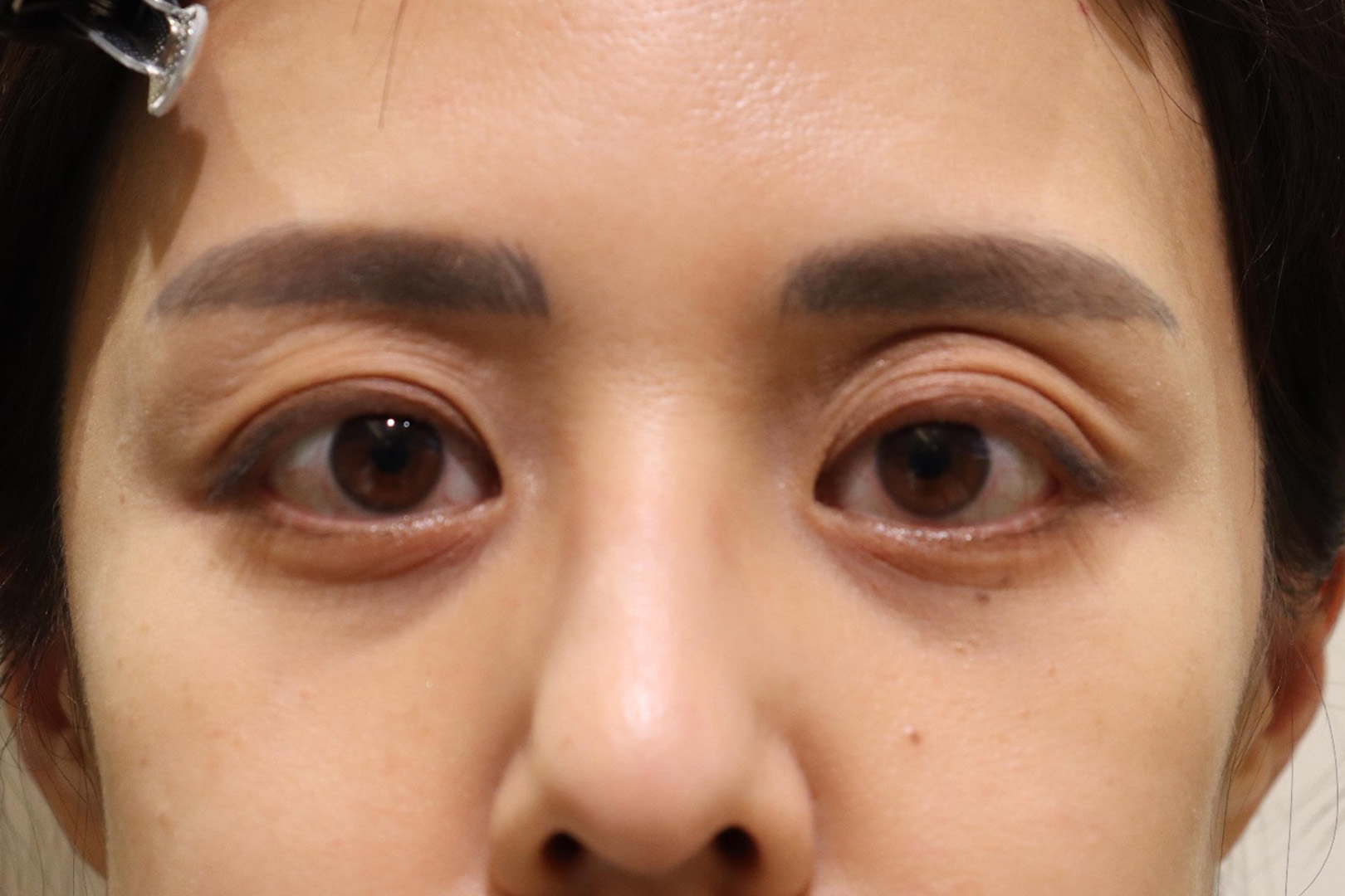
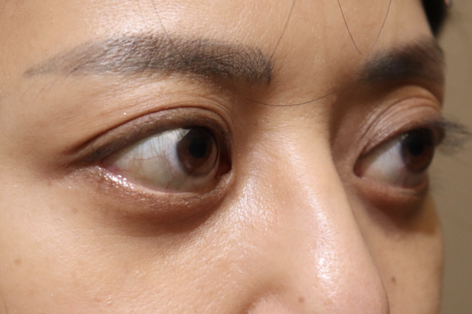
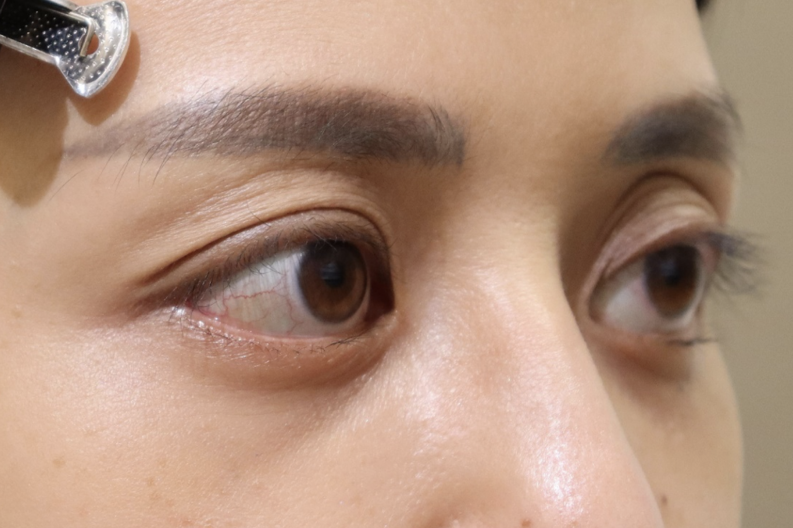
Dark circles under the eyes caused by Thyroid Eye Disease can also be treated.
In our practice, orbital fat removed during orbital decompression surgery is grafted into the dark circles, thereby removing the dark circles under the eyes almost completely. Thyroid eye disease can cause the eyes to appear open, which is called eyelid retraction. Eyelid retraction appears primarily on the upper eyelid, but some patients develop it on the lower eyelid.
This is also slightly improved by orbital decompression, but can be further improved by fat grafting.
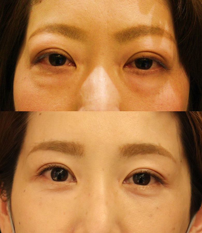
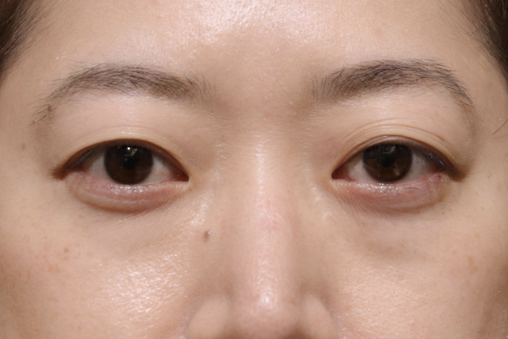
Cases of Thyroid Eye Disease
Case.1
Pre-Illness
Post-Illness
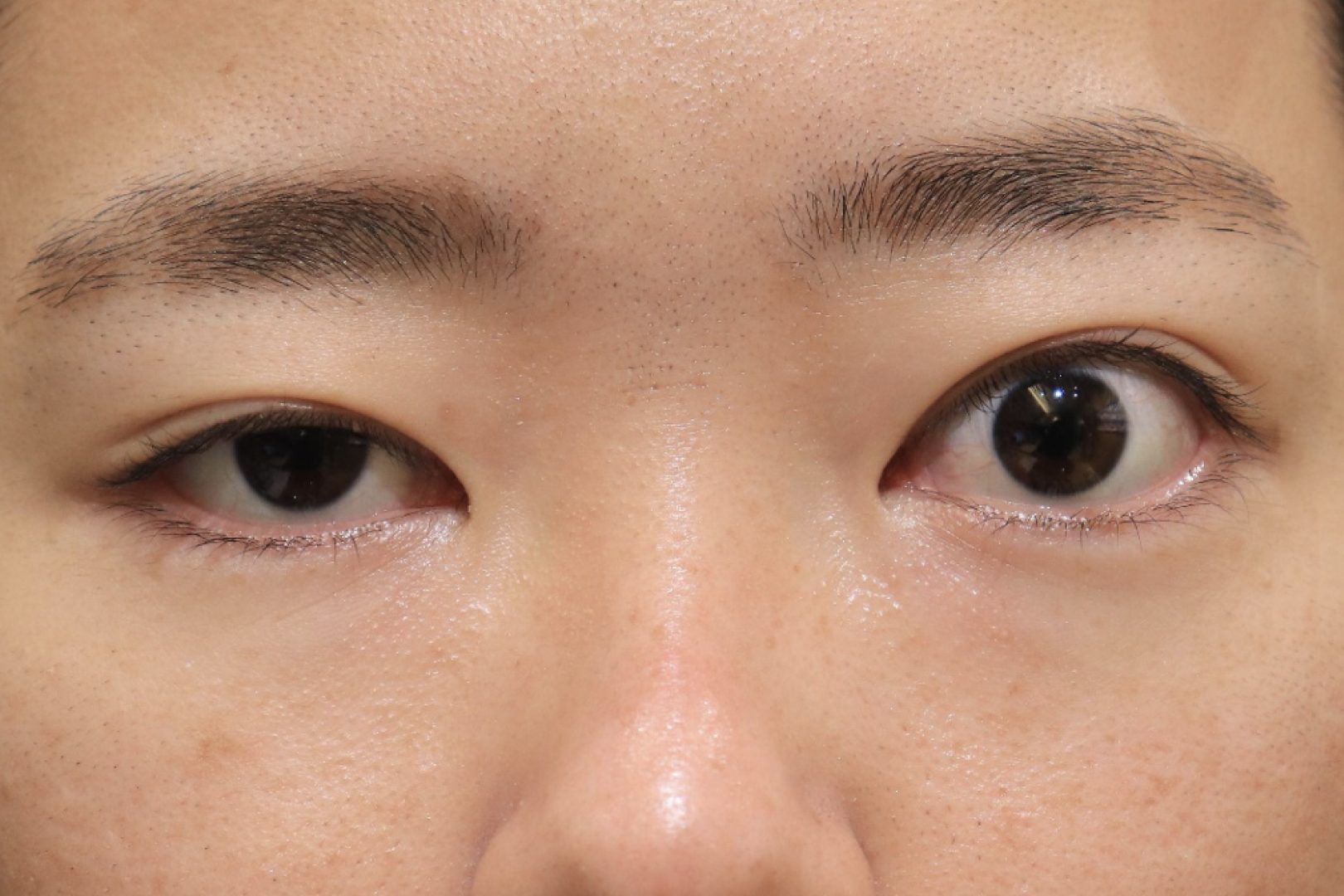
A comparison of before and after the onset of thyroid eye disease shows a significant change in facial appearance. The changes occur in both the upper and lower eyelids. The result is a completely different person.
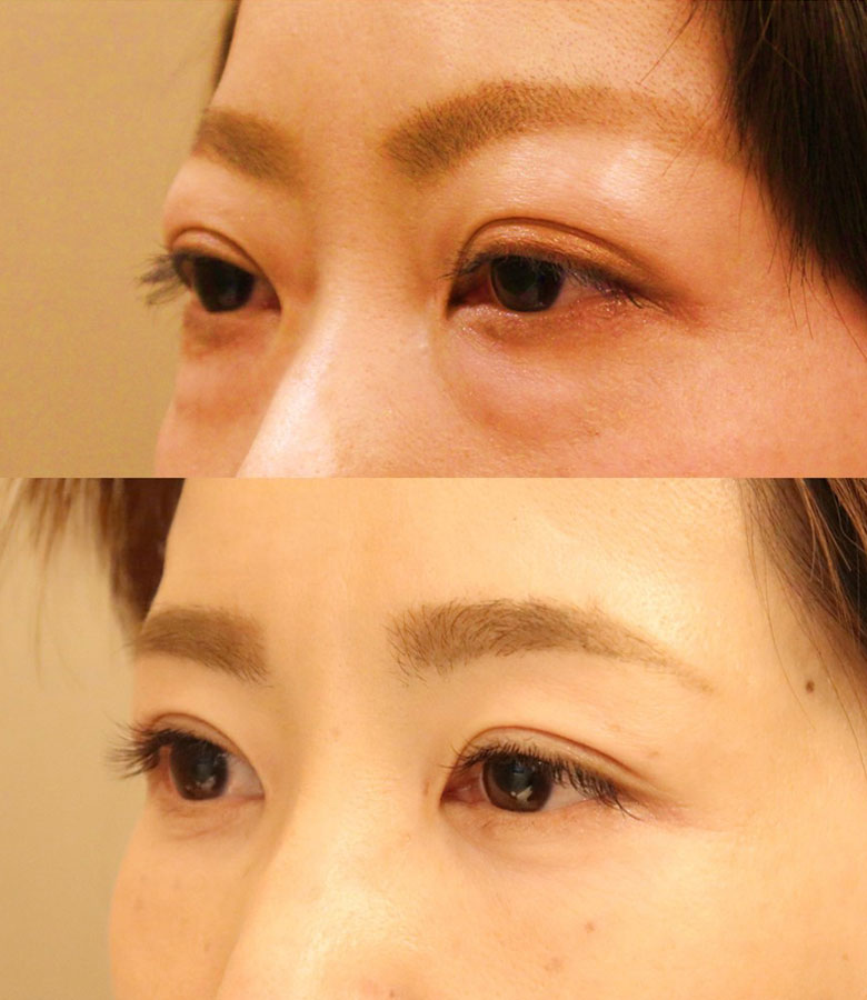
Before surgery
The eyelids are swollen and the eyes are large. Dark circles appear not only on the upper eyelids but also on the lower eyelids. The distance between the eyes also appears to be widened.
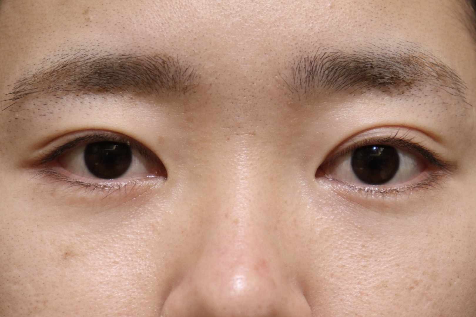
3 months after surgery
The upper eyelid bulge has disappeared and has taken on a natural shape. Dark circles on the lower eyelids have also disappeared and the distance between the eyes has narrowed.
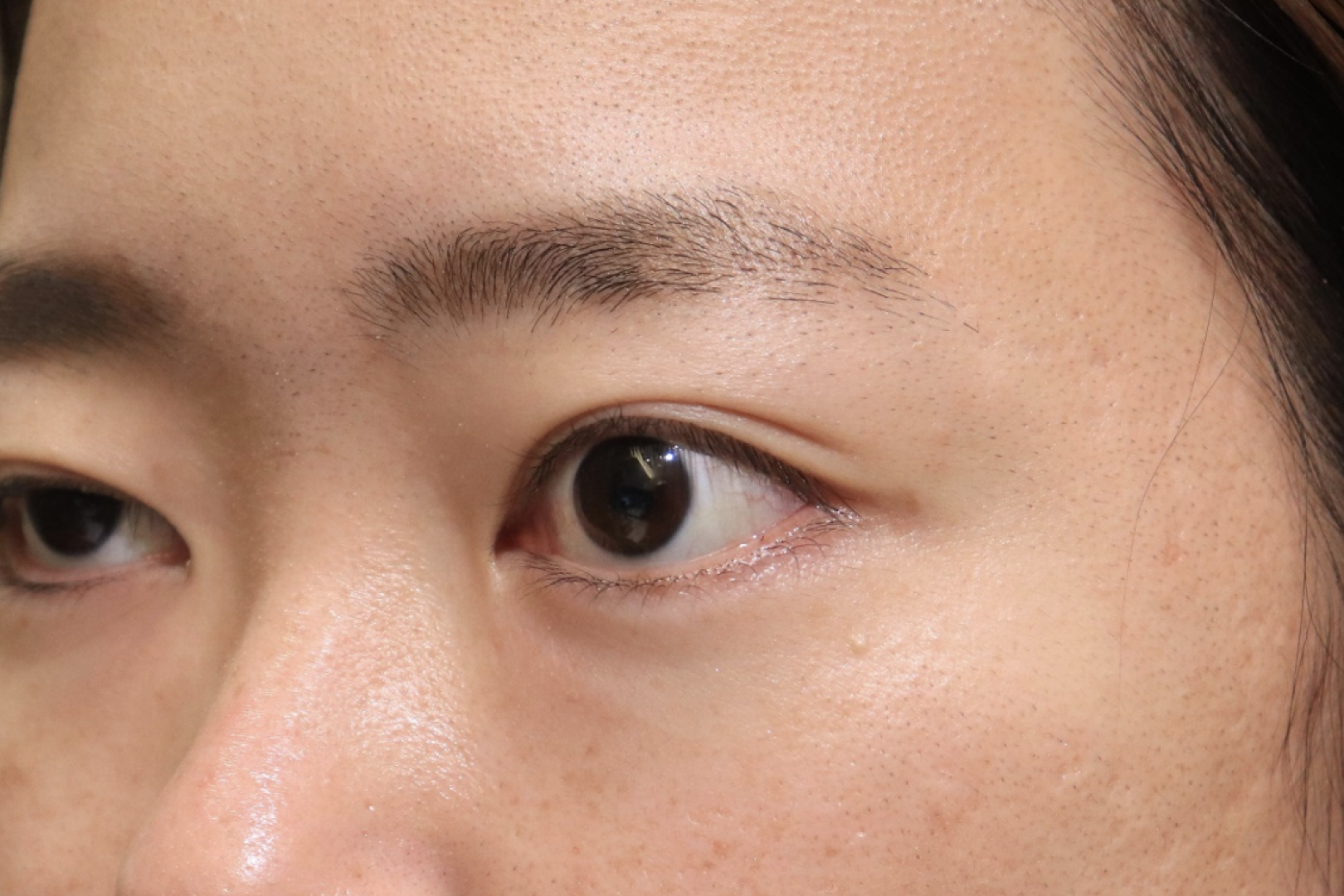
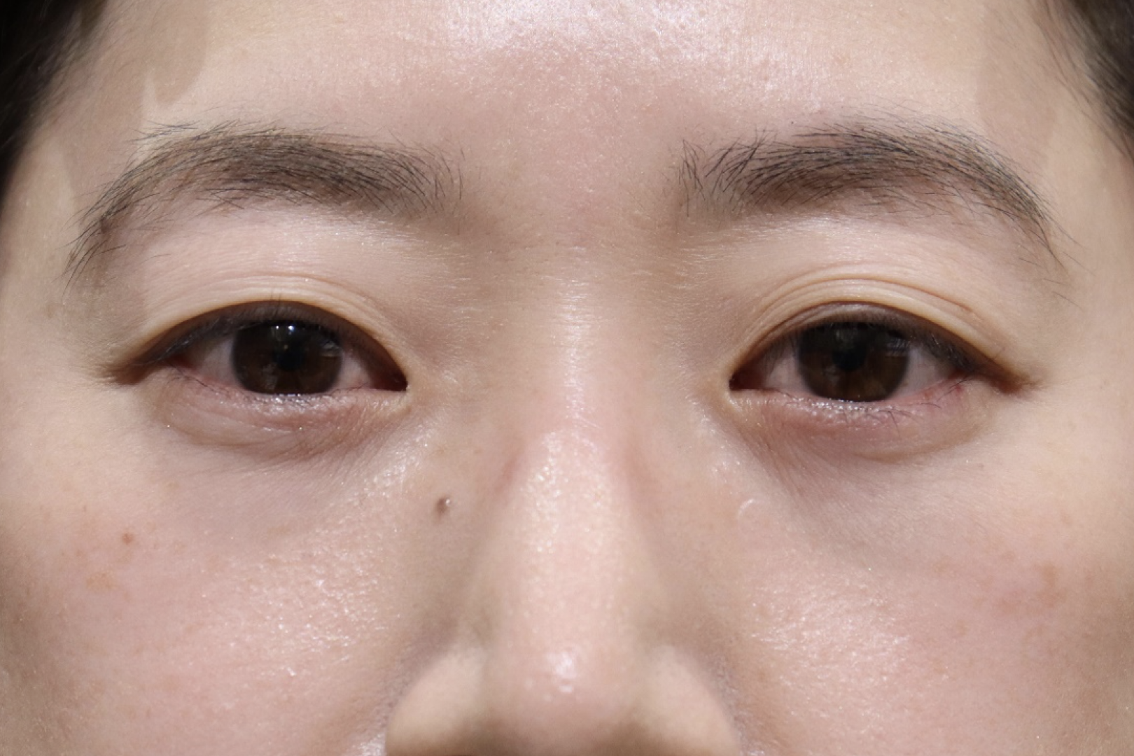
Before surgery
The eyelids are swollen and the eyes are large, giving the appearance of staring. Dark circles appear on the lower eyelids as well as the upper eyelids.
3 months after surgery
The upper eyelid bulge has disappeared and taken on a natural shape. Dark circles on the lower eyelids have also disappeared.
Chief complaint
Thyroid eye disease
Treatment cost
Dr. Kashima: 1,980,000 yen
Dr. Yamana, Dr. Kikuchi, Dr. Kotaki: 880,000 yen
Other than the above *No designated surgeon (monitor required): 440,000 yen
Fat grafting to dark circles under the eyes +220,000 yen
Treatment content
Orbital decompression
Risks
Bleeding, swelling, new or worsening diplopia, vision loss, overcorrection, undercorrection
Case.2
Pre-Illness
Post-Illness
A comparison of before and after the onset of thyroid eye disease shows a significant change in facial appearance. The changes occur in both the upper and lower eyelids. The result is a completely different person.
Before surgery
The eyelids are swollen and the eyes are large. Dark circles appear not only on the upper eyelids but also on the lower eyelids. The distance between the eyes also appears to be widened.
3 months after surgery
The upper eyelid bulge has disappeared and natural shape has been achieved. Dark circles on the lower eyelids have also disappeared and the distance between the eyes has narrowed.
Before surgery
The eyelids are swollen and the eyes are large. Dark circles appear on the lower eyelids as well as the upper eyelids.
3 months after surgery
The bulge in the upper eyelid has disappeared and a natural shape has been achieved. The change is easy to see when looking at the right eye. Dark circles on the lower eyelid have also disappeared.
Chief complaint
Thyroid eye disease
Treatment cost
Dr. Kashima: 1,980,000 yen
Dr. Yamana, Dr. Kikuchi, Dr. Kotaki: 880,000 yen
Other than the above *No designated surgeon (monitor required): 440,000 yen
Fat grafting to dark circles under the eyes +220,000 yen
Treatment content
Orbital decompression
Risks
Bleeding, swelling, new or worsening diplopia, vision loss, overcorrection, undercorrection
Case.3
Before surgery
The eyelids are swollen and the eyes are large.
3 months after surgery
The upper eyelid bulge has disappeared and taken on a natural shape. Dark circles on the lower eyelids have also disappeared.
Before surgery
まThe lid is swollen. Dark circles appear on the lower eyelids as well as the upper eyelids.
3 months after surgery
上The eyelid bulge has disappeared and a natural shape has been achieved. It is especially easy to compare the change in the left eye on the back side. Dark circles on the lower eyelid have also disappeared.
Chief complaint
Thyroid eye disease
Treatment cost
Dr. Kashima: 1,980,000 yen
Dr. Yamana, Dr. Kikuchi, Dr. Kotaki: 880,000 yen
Other than the above *No designated surgeon (monitor required): 440,000 yen
Fat grafting to dark circles under the eyes +220,000 yen
Treatment content
Orbital decompression
Risks
Bleeding, swelling, new or worsening diplopia, vision loss, overcorrection, undercorrection
Case.4
Before surgery
The eyelids are swollen, especially the left eye is wide open. The distance between the eyes also seems to be widening.
3 months after surgery
The upper eyelid bulge has disappeared, and a natural shape has been achieved. The eye area has also been improved. Dark circles on the lower eyelids have also disappeared.
Before surgery
The eyelids are swollen and the eyes are large. Dark circles appear not only on the upper eyelids but also on the lower eyelids. The distance between the eyes also appears to be widened.
3 months after surgery
The upper eyelid bulge has disappeared and has taken on a natural shape. Dark circles on the lower eyelids have also disappeared and the distance between the eyes has narrowed.
Chief complaint
Thyroid eye disease
Treatment cost
Dr. Kashima: 1,980,000 yen
Dr. Yamana, Dr. Kikuchi, Dr. Kotaki: 880,000 yen
Other than the above *No designated surgeon (monitor required): 440,000 yen
Fat grafting to dark circles under the eyes +220,000 yen
Treatment content
Orbital decompression
Risks
Bleeding, swelling, new or worsening diplopia, vision loss, overcorrection, undercorrection
Case.5
Pre-Illness
Post-Illness
A comparison of before and after the onset of thyroid eye disease shows a change in facial appearance. The changes occur in both the upper and lower eyelids. The result is a completely different person.
Before surgery
The eyelids are swollen and the eyes are large. Dark circles appear not only on the upper eyelids but also on the lower eyelids. The distance between the eyes is also enlarged.
3 months after surgery
The upper eyelid bulge has disappeared and has taken on a natural shape. Because the puffiness has disappeared, fine lines and wrinkles have appeared. Dark circles on the lower eyelids have also disappeared and the distance between the eyes has narrowed.
Before surgery
The eyelids are swollen and the eyes are large. Dark circles appear on the lower eyelids as well as the upper eyelids.
3 months after surgery
The upper eyelid bulge has disappeared and has taken on a natural shape. Dark circles on the lower eyelids have also disappeared and the distance between the eyes has narrowed.
Chief complaint
Thyroid eye disease
Treatment cost
Dr. Kashima: 1,980,000 yen
Dr. Yamana, Dr. Kikuchi, Dr. Kotaki: 880,000 yen
Other than the above *No designated surgeon (monitor required): 440,000 yen
Fat grafting to dark circles under the eyes +220,000 yen
Treatment content
Orbital decompression
Risks
Bleeding, swelling, new or worsening diplopia, vision loss, overcorrection, undercorrection
Case.6
Pre-Illness
Since the eyelids were somewhat concave before the onset of the disease, it can be seen that they became closer to their original facial appearance after the surgery.
Before surgery
The eyelids are swollen and the eyes are large. Dark circles appear not only on the upper eyelids but also on the lower eyelids. The distance between the eyes also appears to be widened.
3 months after surgery
The upper eyelid bulge has disappeared and natural shape has been achieved. Dark circles on the lower eyelids have also disappeared and the distance between the eyes has narrowed.
Before surgery
The eyelids are swollen and the eyes are large. Dark circles appear on the lower eyelids as well as the upper eyelids.
3 months after surgery
The upper eyelid bulge has disappeared and taken on a natural shape. Dark circles on the lower eyelid have also disappeared. The difference can be seen in the right eye at the back.
Chief complaint
Thyroid eye disease
Treatment cost
Dr. Kashima: 1,980,000 yen
Dr. Yamana, Dr. Kikuchi, Dr. Kotaki: 880,000 yen
Other than the above *No designated surgeon (monitor required): 440,000 yen
Fat grafting to dark circles under the eyes +220,000 yen
Treatment content
Orbital decompression
Risks
Bleeding, swelling, new or worsening diplopia, vision loss, overcorrection, undercorrection
Case.7
Pre-Illness
Prior to the onset of the disease, the lower eyelid was in normal position and the whites of the eyes were not exposed.
Before surgery
The eyelids are swollen, indicating large eyes. The lower eyelids are pulled back, exposing the whites of the eyes under the black eyes.
3 months after surgery
The upper eyelid bulge has disappeared and a natural shape has been achieved. The distance between the eyes has also narrowed. The position of the lower eyelid has also improved and the whites of the eyes are less exposed.
Before surgery
The eyelids are swollen, indicating large eyes. The lower eyelids are pulled back, exposing the whites of the eyes under the black eyes.
3 months after surgery
The upper eyelid bulge has disappeared and taken on a more natural shape. The position of the lower eyelid has also improved and the whites of the eyes are less exposed.
Chief complaint
Thyroid eye disease
Treatment cost
Dr. Kashima: 1,980,000 yen
Dr. Yamana, Dr. Kikuchi, Dr. Kotaki: 880,000 yen
Other than the above *No designated surgeon (monitor required): 440,000 yen
Fat grafting to dark circles under the eyes +220,000 yen
Treatment content
Orbital decompression
Risks
Bleeding, swelling, new or worsening diplopia, vision loss, overcorrection, undercorrection
Case.8
Pre-Illness
It can be seen that the eye originally had a concave upper eyelid before the onset of thyroid eye disease.
Before surgery
The eyelids are swollen and the eyes are large. Dark circles appear not only on the upper eyelids but also on the lower eyelids. The distance between the eyes also appears to be widened.
3 months after surgery
The upper eyelid bulge has disappeared and is slightly concave.Dark circles on the lower eyelids have also disappeared and the distance between the eyes has narrowed.
Before surgery
The eyelids are swollen and the eyes are large. Dark circles appear not only on the upper eyelids but also on the lower eyelids. The distance between the eyes also appears to be widened.
3 months after surgery
The upper eyelid bulge has disappeared and is slightly concave. Dark circles on the lower eyelids have also disappeared.
Chief complaint
Thyroid eye disease
Treatment cost
Dr. Kashima: 1,980,000 yen
Dr. Yamana, Dr. Kikuchi, Dr. Kotaki: 880,000 yen
Other than the above *No designated surgeon (monitor required): 440,000 yen
Fat grafting to dark circles under the eyes +220,000 yen
Treatment content
Orbital decompression
Risks
Bleeding, swelling, new or worsening diplopia, vision loss, overcorrection, undercorrection
Case.9
Pre-Illness
This is before the onset of thyroid eye disease. If the disease developed in the teenage years, we do not know the original facial appearance, so we match it to what it would have looked like had it grown up.
Before surgery
The eyelids are swollen and the eyes are large. Dark circles appear not only on the upper eyelids but also on the lower eyelids. The distance between the eyes also appears to be widened.
3 months after surgery
上The eyelid bulges have disappeared and have become slightly concave. Dark circles on the lower eyelids have also disappeared and the distance between the eyes has narrowed and become more balanced.
Before surgery
The eyelids are swollen and the eyes are large. Dark circles appear not only on the upper eyelids but also on the lower eyelids. The distance between the eyes also appears to be widened.
3 months after surgery
The upper eyelid bulge has disappeared and is slightly concave. Dark circles on the lower eyelids have also disappeared and the distance between the eyes has been narrowed and balanced.
Chief complaint
Thyroid eye disease
Treatment cost
Dr. Kashima: 1,980,000 yen
Dr. Yamana, Dr. Kikuchi, Dr. Kotaki: 880,000 yen
Other than the above *No designated surgeon (monitor required): 440,000 yen
Fat grafting to dark circles under the eyes +220,000 yen
Treatment content
Orbital decompression
Risks
Bleeding, swelling, new or worsening diplopia, vision loss, overcorrection, undercorrection
Case.10
Before surgery
The eyelids are swollen and the eyes are large. The distance between the eyes also seems to be widened.
3 months after surgery
The upper eyelid bulge has disappeared and is slightly concave. The distance between the eyes has also been narrowed and balanced.
Before surgery
The eyelids are swollen and the eyes are large. The distance between the eyes also seems to be widened.
3 months after surgery
The upper eyelid bulge has disappeared and is now slightly concave. The distance between the eyes has also narrowed and become more balanced. The difference can be seen when looking at the left eye in the back.
Chief complaint
Thyroid eye disease
Treatment cost
Dr. Kashima: 1,980,000 yen
Dr. Yamana, Dr. Kikuchi, Dr. Kotaki: 880,000 yen
Other than the above *No designated surgeon (monitor required): 440,000 yen
Fat grafting to dark circles under the eyes +220,000 yen
Treatment content
Orbital decompression
Risks
Bleeding, swelling, new or worsening diplopia, vision loss, overcorrection, undercorrection
Case.11
Chief complaint
Thyroid eye disease
Treatment cost
Dr. Kashima: 1,980,000 yen
Dr. Yamana, Dr. Kikuchi, Dr. Kotaki: 880,000 yen
Other than the above *No designated surgeon (monitor required): 440,000 yen
Fat grafting to dark circles under the eyes +220,000 yen
Treatment content
Orbital decompression
Risks
Bleeding, swelling, new or worsening diplopia, vision loss, overcorrection, undercorrection
Orbital Decompression for people without Graves’ disease
Effective for other than Graves’ disease
Orbital decompression can also be performed for eye protrusions that are not caused by Graves’ disease.
If the protruding eyeball causes disfigurement, rabbit’s eye or dry eye, surgery is indicated.
Available for revision surgery after surgery at other hospitals
You may notice that the upper eyelid bulge is improved and the double eyelid is deeper after orbital decompression.
We can also handle cases such as failures at other clinics and cases that need to be redone, so if you have any problems, please come in for a consultation first.
Estimated treatment fees
both eyes/ one eye
Orbital decompression
20,000USD (Included coordinator fee)
Special price Surgery fee*1
20%OFF
*1:We offer treatment at a discounted rate for those willing to cooperate with us by allowing photography before and after the procedure, as well as video recording during the surgery, for publication on our website.
Risks associated with this treatment and their incidence
Internal bleeding may occur at the wound site due to surgery. At first, internal bleeding looks like a red bruise, but it turns yellow and moves downward under the skin due to gravity, disappearing in about 3 weeks.
The swelling will mostly subside within the first two weeks.
It takes about six months for complete disappearance. If a hematoma forms in the wound, surgery is required to remove it.
If there is no diplopia before surgery, eye movement disorders will appear after surgery, resulting in double vision. It often disappears by the next day, but it may remain.
If diplopia remains, it will gradually improve over 3 to 6 months, but diplopia remains in 0-3% of cases with lipectomy alone, 3-6% with lateral walls, and 10-65% with medial walls. It is said that then. In that case, strabismus surgery may be necessary. Even if double vision does not appear in frontal vision, there is a 10% chance that double vision will remain in peripheral vision (up, down, left, and right).
After the surgery, your pupils may dilate or it may become difficult to see up close, but this will gradually improve over about six months.
If damage occurs to the important nerves related to the eye or the blood vessels that feed them, vision and visual field problems may occur, leading to blindness. The incidence is approximately 1%, including mild cases.
Since the surgery is performed close to the brain, infection can lead to serious conditions.
The scar will gradually become less noticeable after surgery, but it may become noticeable or become a keloid. If the wound disintegrates after surgery, suturing will be required again. Orbital cellulitis may occur due to infection.
Consent form text
To patients and their families undergoing decompression surgery for thyroid eye disease
This manual explains the orbital decompression surgery for thyroid eye disease. If you have any questions, please ask your doctor. If you are undergoing treatment, please read the “Consent form PDF” below and sign it.
Member of the American Society of Ophthalmic Plastic and Reconstructive Surgery (ASOPRS)
International and Domestic Activities
2009
Singapore National Eye Center
2010
Asia-Pacific Society of Ophthalmic Plastic Surgery in Beijing Invited Speaker
2011
European Society of Ophthalmic Plastic Surgery in Como, Italy
2012
World Ophthalmology Congress in Abu Dhabi Invited Speaker Asia ARVO
Singapore Invited Speaker APAO Busan Invited Speaker
American Academy of Ophthalmology Chicago Invited Speaker
2013
APAO Hyderabad Invited Speaker American Pediatric Ophthalmology Society Singapore Invited Speaker European Society of Ophthalmic Plastic Surgery Barcelona
2014
World Ophthalmology Congress Tokyo Invited Speaker Asia-Pacific Society of Ophthalmic Plastic Surgery Delhi American Society of Ophthalmic Plastic and Reconstructive Surgery Chicago
2015
APAO Guangzhou Invited Speaker
2016
KSAS (Korea Society of Aesthetic Surgery) meeting in Seoul ITEDS (International Thyroid Eye Disease) meeting in London AP SOPRS & JSOPRS joint meeting session chair iseminer Numerous lectures on surgical treatment of thyroid eye disease.
2017
American Academy of Ophthalmology instructor, Korean Society of Oculoplastic Surgery invited speaker, Chinese Society of Oculoplastic Surgery invited speaker
2018
Asia-Pacific International Society APAO invited speaker
2019
Asia-Pacific International Society APAO invited speaker, ITEDS invited speaker, OPAIC invited speaker
2021
Asia-Pacific Society of Ophthalmic Plastic Surgery invited speaker
2023
Asia Pacific Academy of Ophthalmology (Kuala Lumpur)
Oculoplastic Lecture (Bangkok)
Asia International Thyroid Eye Disease (Singapore)
World Society of Ophthalmic Plastic and Reconstructive Surgery (Dubai)
UCLA cadaver course (Los Angeles)
Thai Society of Ophthalmic Plastic and Reconstructive Surgery (Bangkok)
European Society of Ophthalmic Plastic and Reconstructive Surgery (Napoli)
Oculoplastic Academy (Coimbra)
American Society of Ophthalmic Plastic and Reconstructive Surgery (San Francisco)
Supervision & Authorship
Supervision
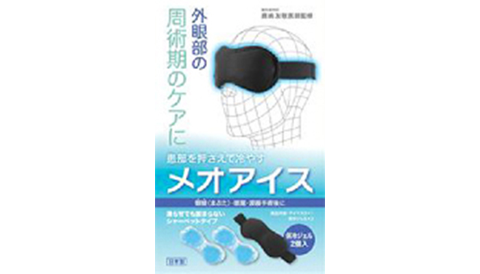
Perioperative care of the external eye
Meo Ice
A medical product developed by Nagoya Glasses to prevent “swelling” and “pain” after surgery around the eyelids.
Authorship
Chief Editor
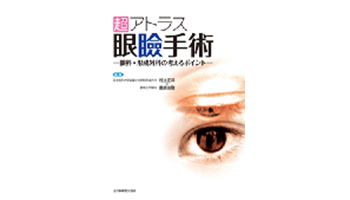
Ultimate Atlas of Eyelid Surgery
An ambitious atlas aiming for collaboration between ophthalmology and plastic surgery, published by All Japan Hospital Publishing. It is clearly explained with continuous color photos and detailed schematics.
Authorship
Chief Editor
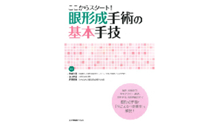
Start Here! Basic Techniques in Eyelid Plastic Surgery
An introductory book for doctors aspiring to specialize in oculoplastic surgery, published by All Japan Hospital Publishing. It thoroughly explains surgical tools and techniques.