Orbital fat decompression for thyroid eye disease and congenital exophthalmos
Minimally invasive orbital fat decompression surgery
Outpatient surgery
Simultaneous surgery on both eyes
Only excess fat is removed
No incision of the skin
In Graves’ disease and thyroid eye disease, the eyeballs protrude forward due to increased fat behind the eyes or swelling of the muscles that move the eyeballs, leading to significant changes in facial appearance.
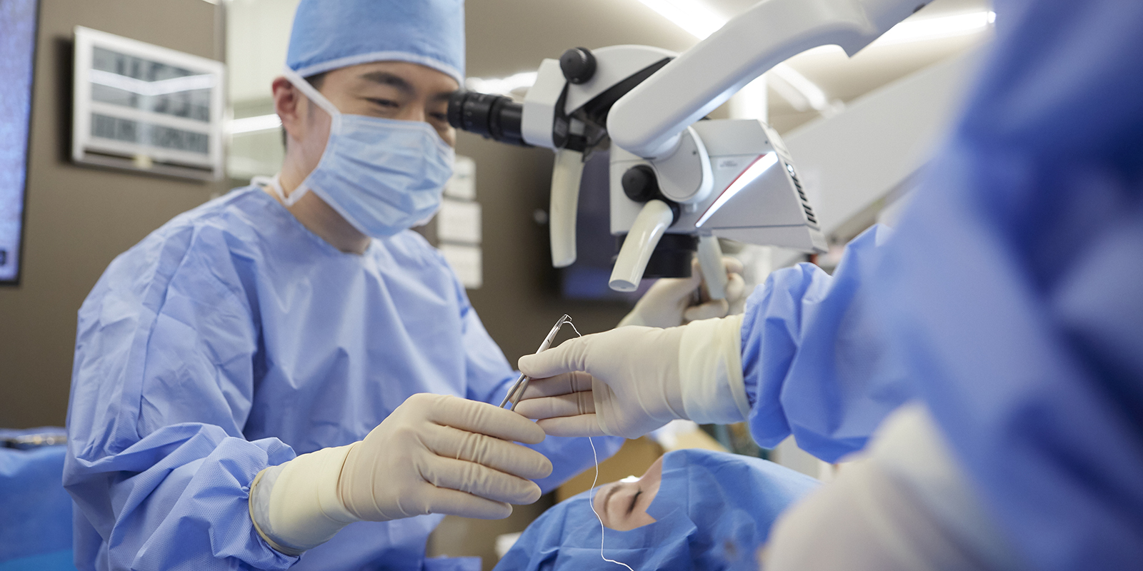
The orbital fat decompression surgery conducted at our clinic is a culmination of global expertise, incorporating cutting-edge medical knowledge acquired through studying in the United States and insights gained from renowned international experts. This surgery is meticulously performed in a Japanese style using microscopes to further minimize damage to essential tissues.
There are no medical institutions worldwide conducting this surgery.
Same-day surgery is possible
This surgery involves removing only the excess fat while avoiding nerves and blood vessels related to visual function, requiring highly delicate and specialized techniques. Decompression surgery involving bone removal was developed because there was no technique to remove fat while avoiding nerves and blood vessels, but it requires significant physical burden due to bone removal, and hospitalization for 1-2 weeks is necessary due to postoperative pain and bleeding.
With our clinic’s orbital fat decompression surgery, both eyes can be operated on simultaneously under general anesthesia on an outpatient basis, allowing patients to return home on the same day. This minimizes the impact on work and home life. Unfortunately, as it is an advanced surgery, it is not covered by health insurance. However, if you are troubled by your condition, we hope you will consider our clinic’s orbital fat decompression surgery.
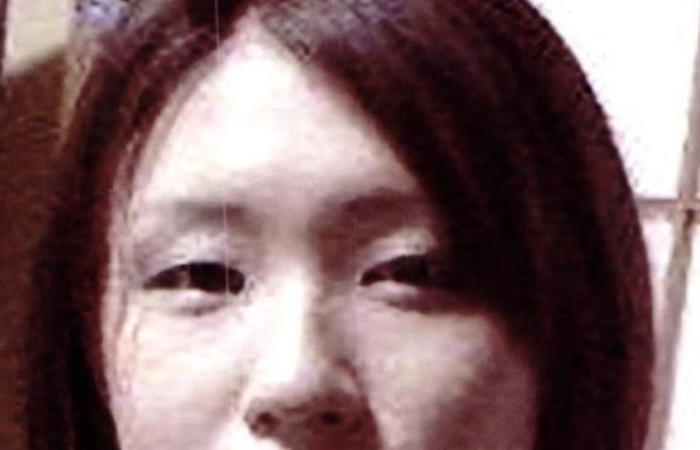
Graves’ disease and thyroid eye disease exhibit significant differences in facial appearance before and after treatment.
This woman underwent the following treatments since the onset of Graves’ ophthalmopathy:
Steroid pulse therapy
Orbital decompression surgery
Additional fat removal
Strabismus surgery (performed at another clinic)
Mainly, the above treatments were administered. When comparing before and after treatment, you can see a significant improvement, and her facial appearance looks completely different.
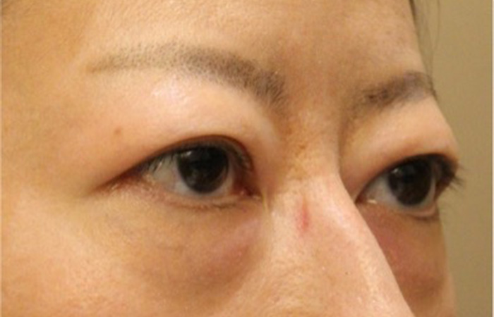
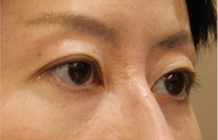
Facial profile
The difference before and after treatment is clear from surgery, and you can regain the beautiful facial features you had before onset.
It seems that Graves' ophthalmopathy was once called the "beautiful woman disease," but looking at this case, it's clear that the idea that "onset makes one beautiful" is a misconception.
What is Graves’ disease in the first place?
A major issue with Graves’ disease and thyroid eye disease is that even if you enter a stable phase after receiving treatments like IVs and oral medications, unlike recovering from a cold, you do not return to normal but end up with a completely different appearance and physique.
The fact that these visual changes predominantly affect young women is particularly problematic. In English, this unsightly bulging of the eyes is called Disfiguring Proptosis.
There are also misunderstandings, such as “the bulging will resolve once the internal medical values stabilize,” but once it has protruded, it does not return to its original state.
Can Graves’ disease and thyroid eye disease be cured?
Many people suffer from these conditions, but there are only a few places nationwide that perform surgical treatments for Graves’ disease and thyroid eye disease.
Moreover, because the surgeries can be quite extensive within the field of ophthalmology, some facilities require several weeks of hospitalization. For this reason, there are hardly any facilities that perform surgery for relatively mild cases of eye bulging.
For such patients, we perform surgeries with a focus on cosmetic perspectives as much as possible, based on our specialized knowledge and experience.
Graves’ disease and thyroid eye disease video collection.
Graves’ disease and thyroid eye disease explanatory video.
What kind of disease is Graves’ ophthalmopathy?? Doctor explains Graves’ ophthalmopathy!
Assessment of activity in Graves’ ophthalmopathy About the active and inactive phases ~ Doctor explains Graves’ ophthalmopathy!
Treatment of active phase of Graves’ ophthalmopathy About the method of steroid administration~ Doctor explains Graves’ ophthalmopathy!
Autoantibodies in Graves’ ophthalmopathy: TRAb/TSAb explained Doctor explains Graves’ ophthalmopathy!
Graves’ disease and thyroid eye disease surgical treatment video.
Graves’ disease and thyroid eye disease explanatory video.
Orbital decompression s urgery treatment video.
Doctor explains Graves’ ophthalmopathy! Oculofacial Clinic Tokyo Dr. Tomoyuki Kashima
Our hospital strives to perform surgeries that minimize the burden on patients, such as post-operative pain and scars, more than any other medical institution in Japan. Please do not hesitate to contact us.
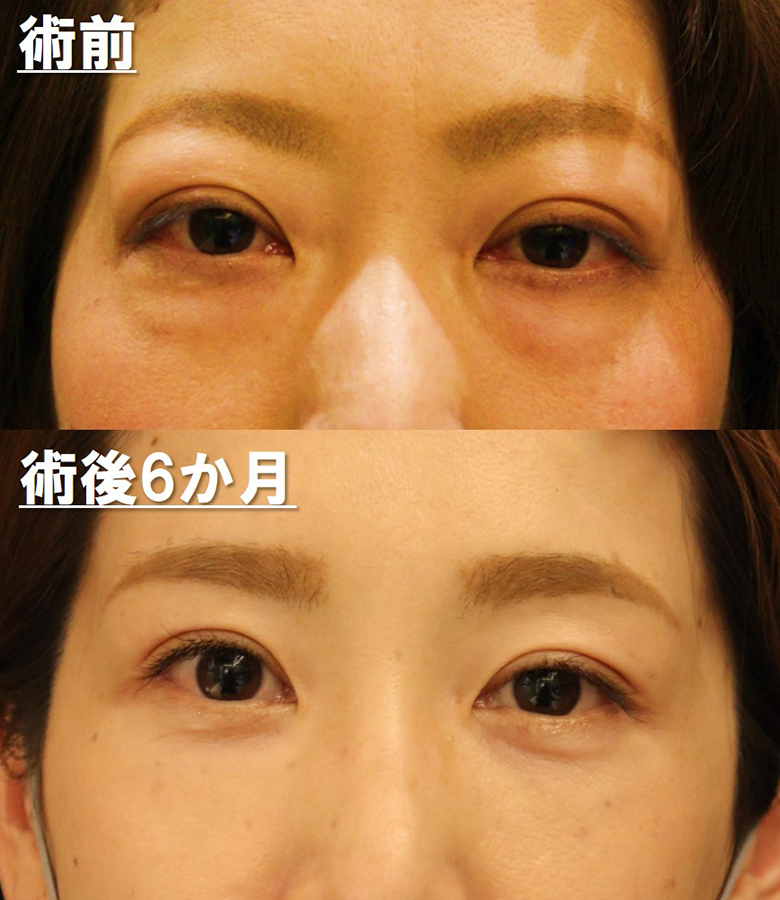
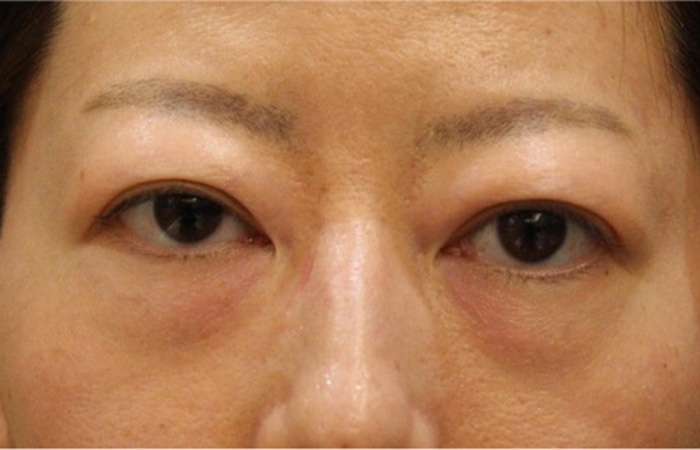
She has fatty hyperplasia of both upper and lower eyelids with ocular protrusion.
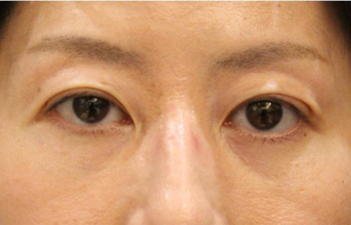
Post
Both upper and lower eyelids show improvement in adipogenesis.There seem to be no fat gain due to thyroid eye disease.
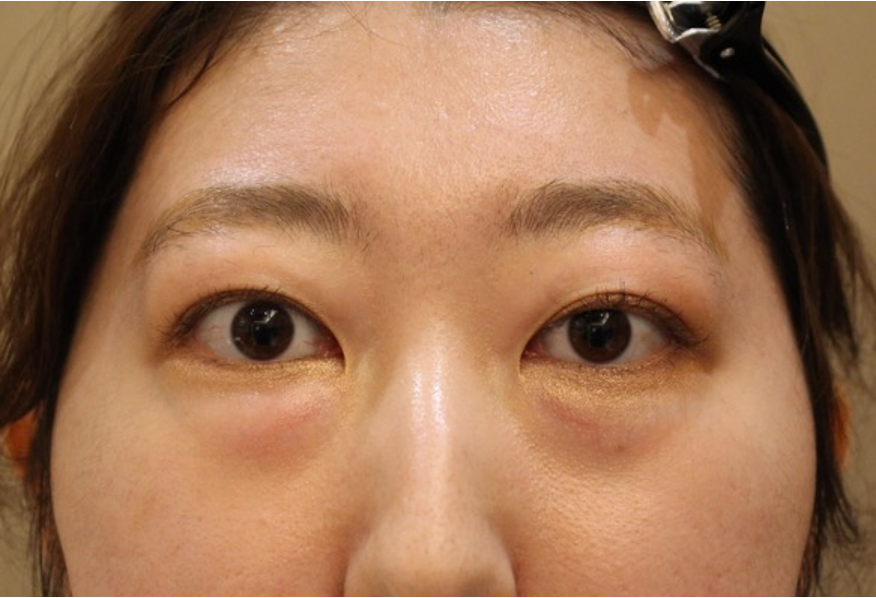
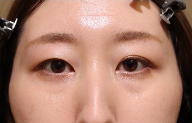
40 years old Female
This is the result after fat decompression. We performed both upper and lower fat decompression for this case.
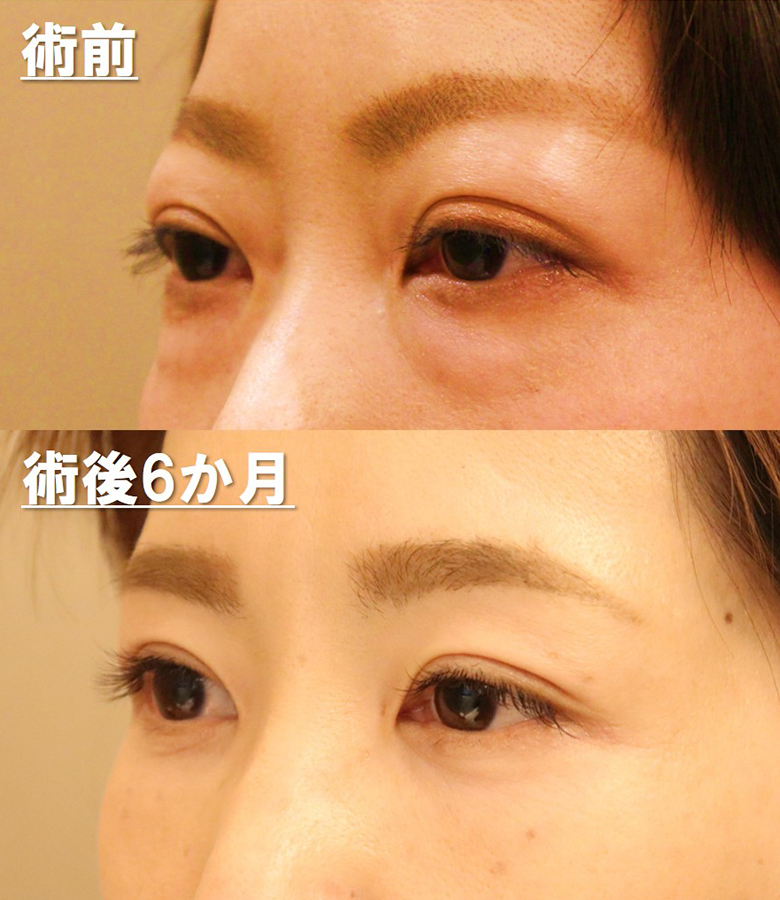
Pre
Before surgery, upper and lower eyelid was puffy and swollen.
Post
After surgery, the eyelid become thin, skinny.Especially, please check far side of upper eyelid, eyelid become sharp and natural.
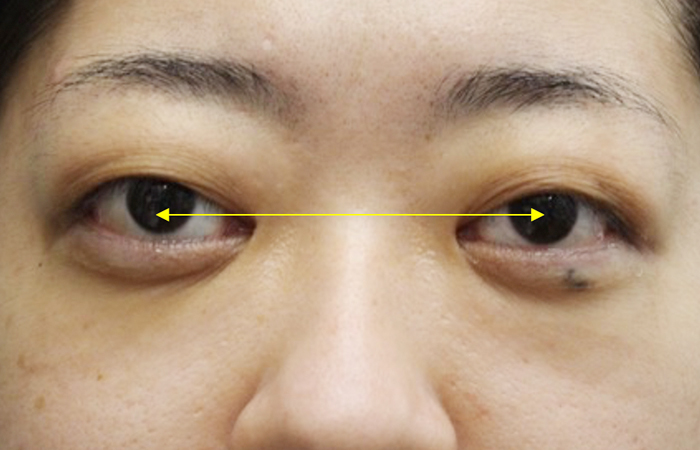
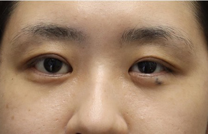
30 years old Female
Pre
After onset of thyroid eye disease, her appearance has completely changed.
Post
After two types of fat decompression, she was back to her old self.Thyroid eye disease make both eyes apart, Removing fat of just behind the lacrimal caruncle, where is medial position of eyeball, eyes become closer.
Treatment with orbital decompression
Proptophthalmia that is not due to Graves’ disease
Orbital decompression can also be performed for congenital proptosis that is not caused by Graves’ disease. People who are born with proptophthalmia, which makes their eyes appear as if they are protruding, do not have protruding eyes, but simply because the bones around their eyes have not developed properly. Very likely.
If tests for Graves' disease show no abnormalities, the patient will be diagnosed with congenital exophthalmia.
At our hospital, we perform a surgery called orbital decompression, which removes the fat and bone behind the eyes, for people with bulging eyes due to Graves' disease.We can apply this technique to treat people with bulging eyes who were born with bulging eyes. We also perform surgery.
However, health insurance cannot cover congenital bulging eyes, so you will have to pay for the entire cost out of pocket. However, if your eyes are too big, there is a good chance that you can make your eyes look more charming by making them smaller and modest. is.
For congenital enophthalmos
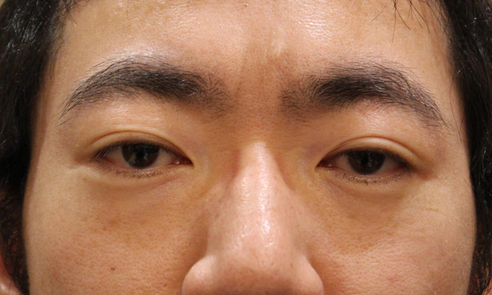
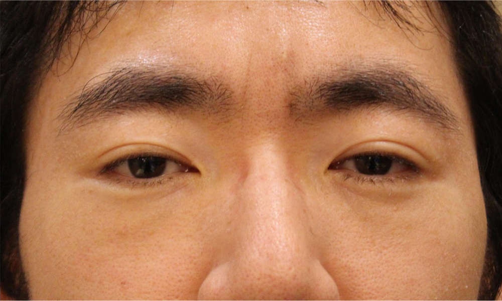
Case1.30 years old Male
Orbital fat decompression can treat congenitally proptotic patient.
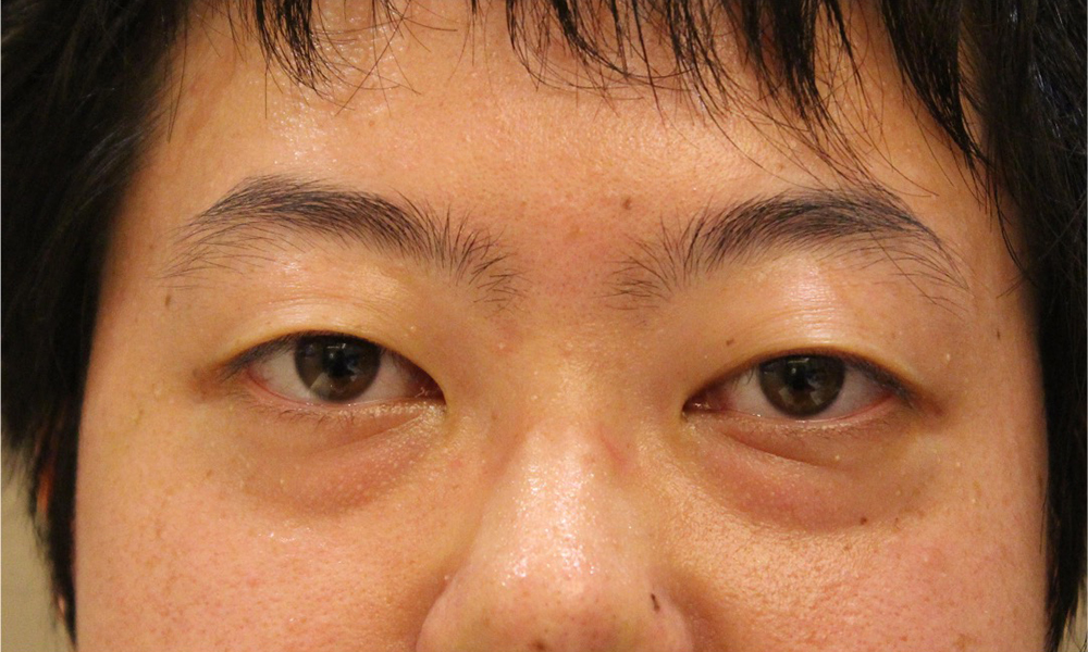
Pre
Her eyes bulge out, her upper eyelids bulge, and her lower eyelids have dark circles.
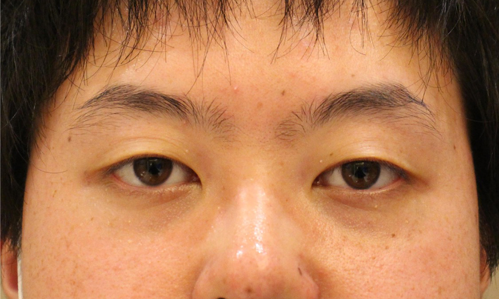
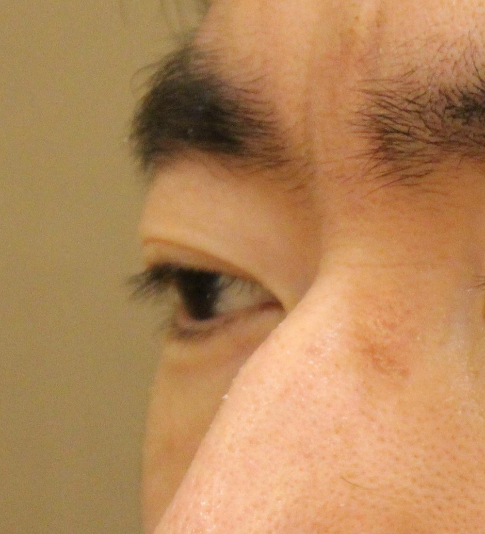
Post
Eyes are smaller, upper eyelid bulge and lower eyelid dark circles have improved.
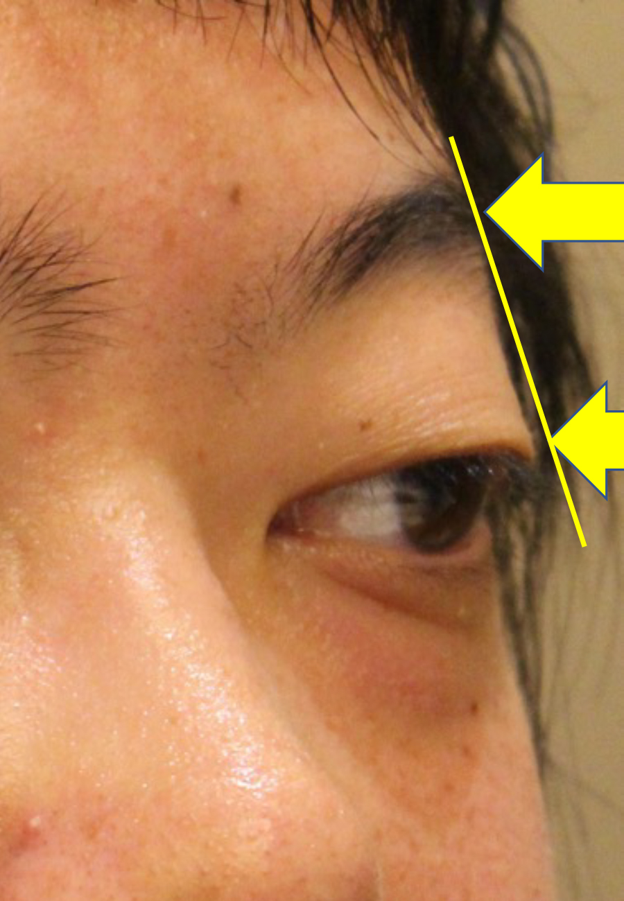
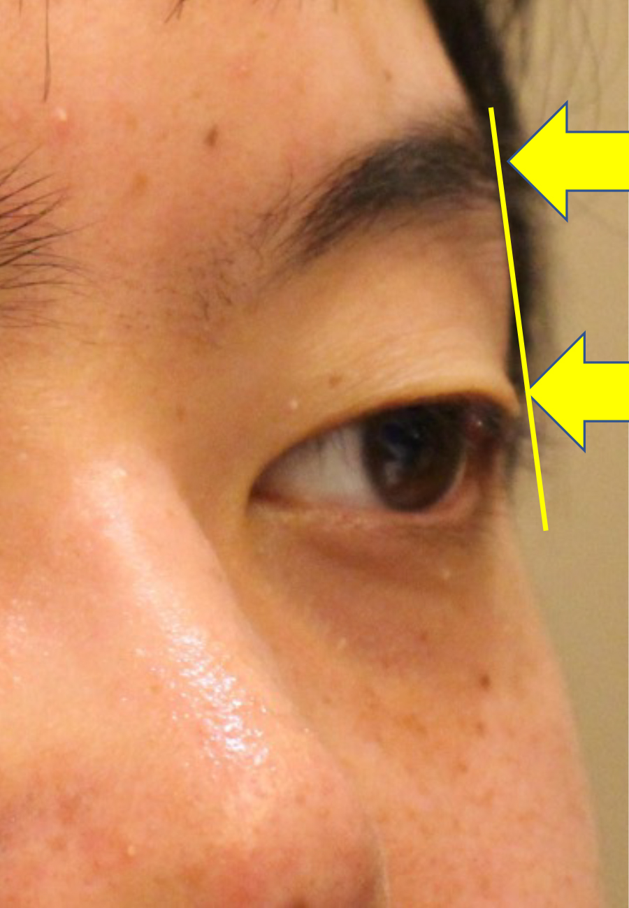
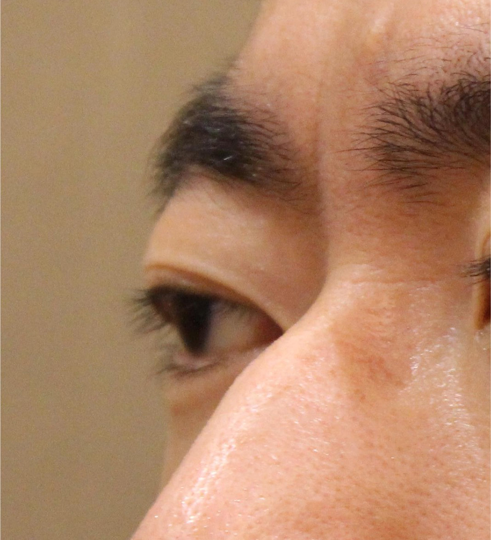
However, from the side, you can understand the change.
Comparing the top of eyelid and eyebrow, it is remarkable eyeball position changed
Different from thyroid eye disease, congenital proptosis do not have increasing volume of fat.
So there is limitation to remove the fat.
Pre
The upper eyelids are swollen and the lower eyelids have dark circles.
Post
Eyes are smaller, upper eyelid bulge and lower eyelid dark circles have improved.
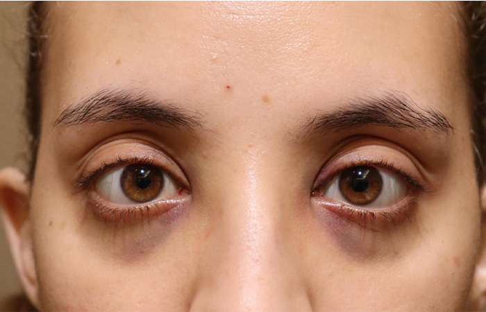
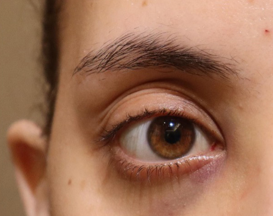
Case2.Explaining orbital decompression using case photos
Compare photos before and after treatment
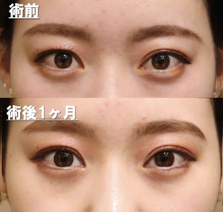
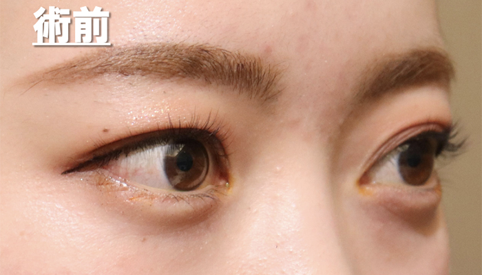
Please take a look at the pre-surgery photos. She has beautiful eyes with double eyelids, but the whites of her eyes are exposed a lot, and her upper eyelids look a little swollen, so she came to our clinic hoping to have these things corrected.
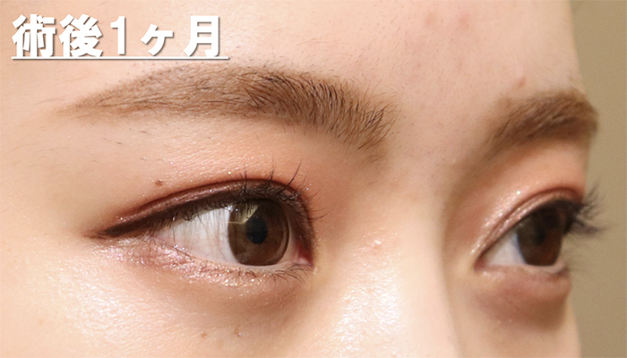
Next, please take a look at the photos taken 1 month after surgery.
There are still traces of bleeding on the lower eyelids and whites of the eyes. It usually takes 3-4 weeks for the bleeding to subside. You can see that the sunken eyes give a gentler impression.
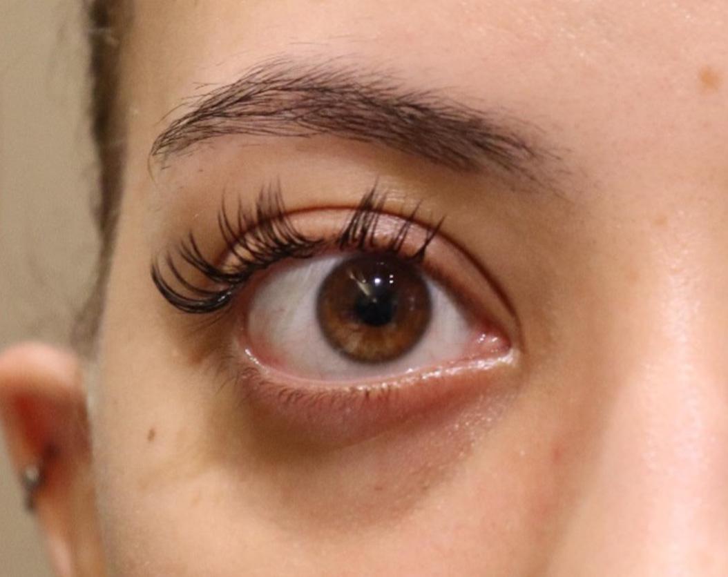
Easy to understand and enlarge
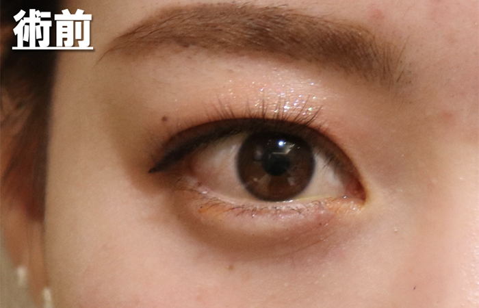
Pre
There is a bulge in the upper eyelids, giving the impression of being a little chubby. The area of the whites of the eyes is also large.
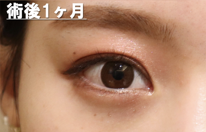
Post
It’s been 2 weeks since the fat was removed from the lower eyelid and behind the eyeball. I still have bleeding in the whites of my eyes and lower eyelids. The area of the whites of the eyes has decreased. Also, because the eyes are sunken, both the upper and lower eyelids cover the eyes, reducing the exposure of brown eyes.
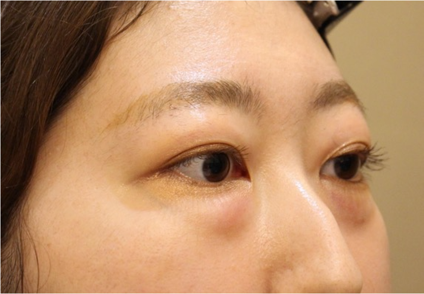
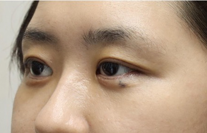
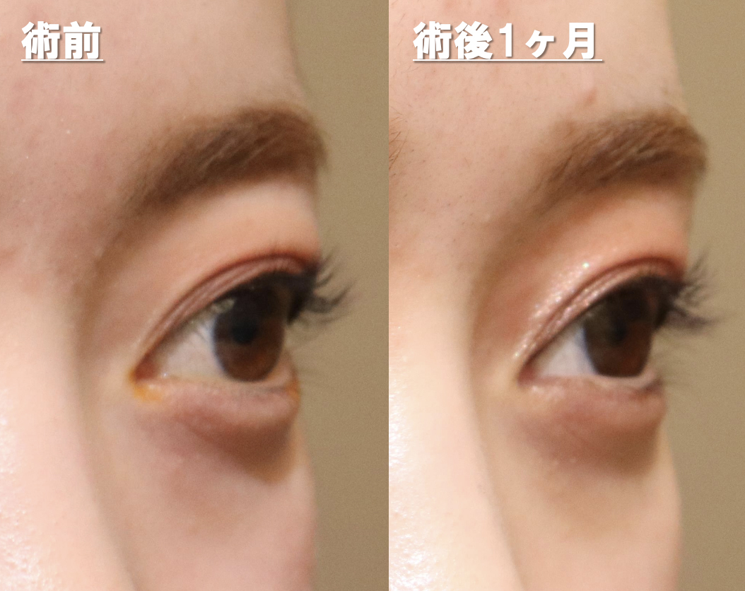
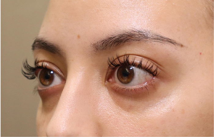
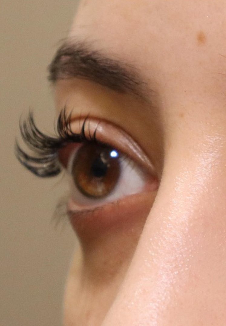
Next is a photo taken from an angle.
Pre
She has beautiful eyes with double eyelids, but the whites of her eyes are exposed a lot, and her upper eyelids have a slightly swollen shape.
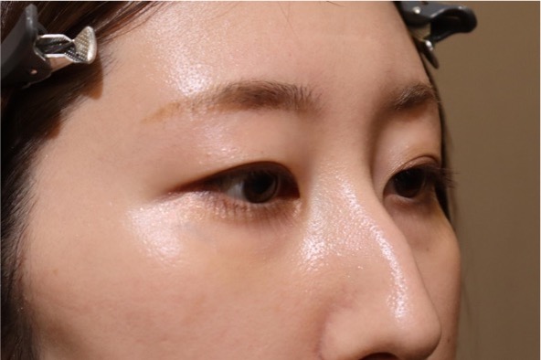
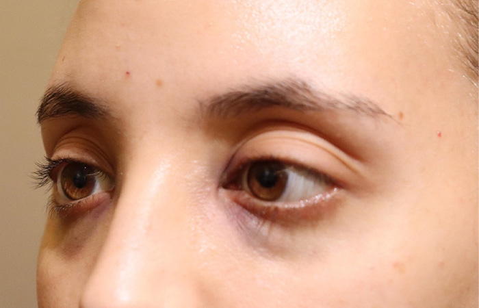
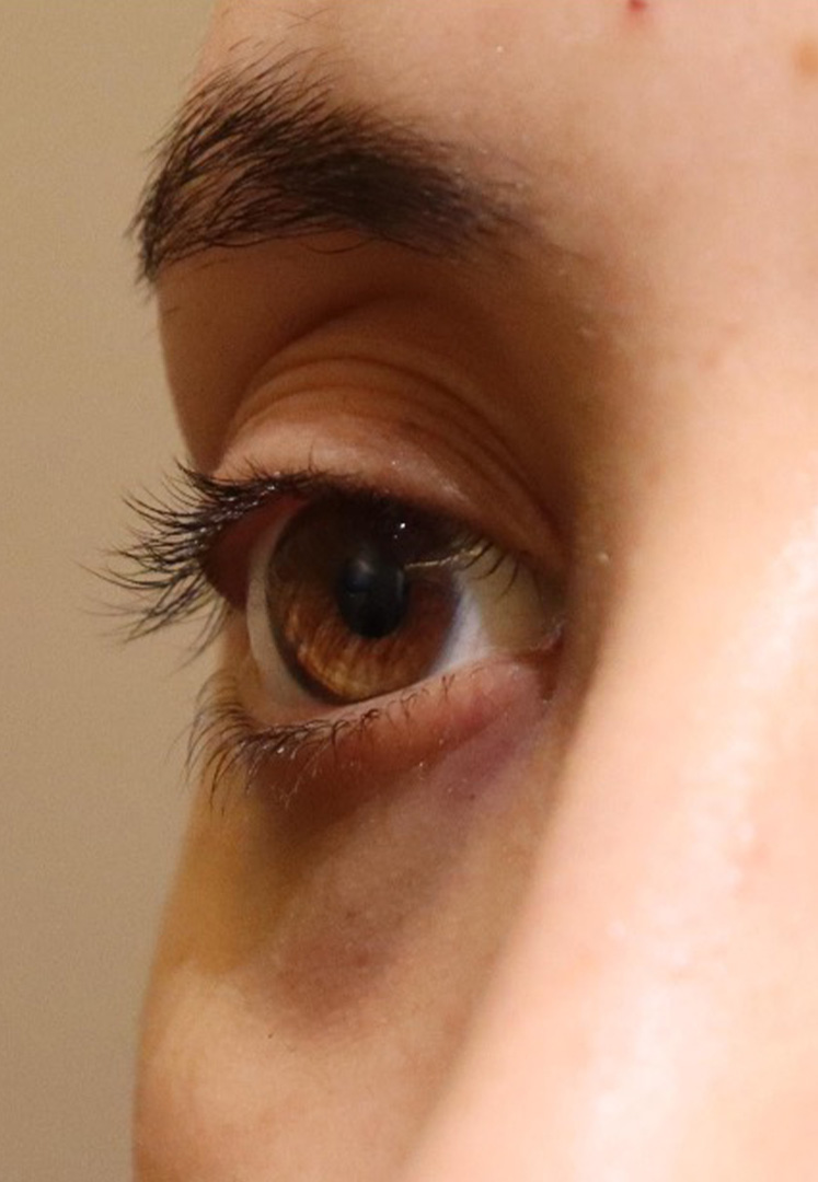
Post
Comparing the area of the whites of the eyes, the area of the whites of the eyes is smaller after surgery. Her eyes are a little smaller, giving her a more beautiful look. My upper eyelids also feel refreshed.
Please pay attention to your left eye
Compare photos before and after treatment
Comparing the left upper eyelid in an oblique photo, you can see that the puffiness has disappeared and the upper eyelid, which used to protrude more than the eyebrows, has become more depressed than the eyebrows after surgery.
In this way, you are born with large eyes (protruding eyes), and our clinic also performs corrections for this. If you have a similar problem, please feel free to contact us.
Risk
Risk of complications
Bleeding, infection, double vision, visual field disturbance, visual impairment, general anesthesia
Cases of congenital bulging eyes
Case3.If the patient need to remove fat more…
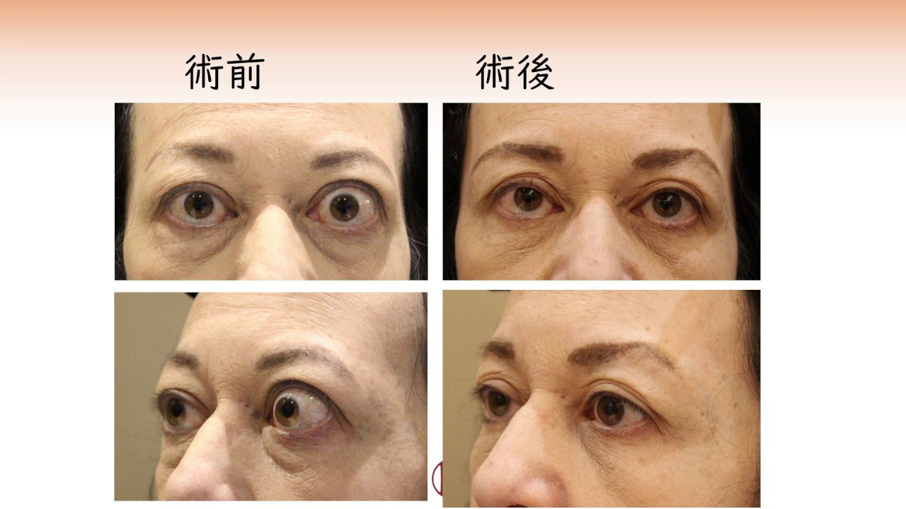
Explaining a case of congenital exophthalmia
Deepening of orbital sulcus cause completely different appearance which may bring impression of aged person.
Compare photos before and after treatment
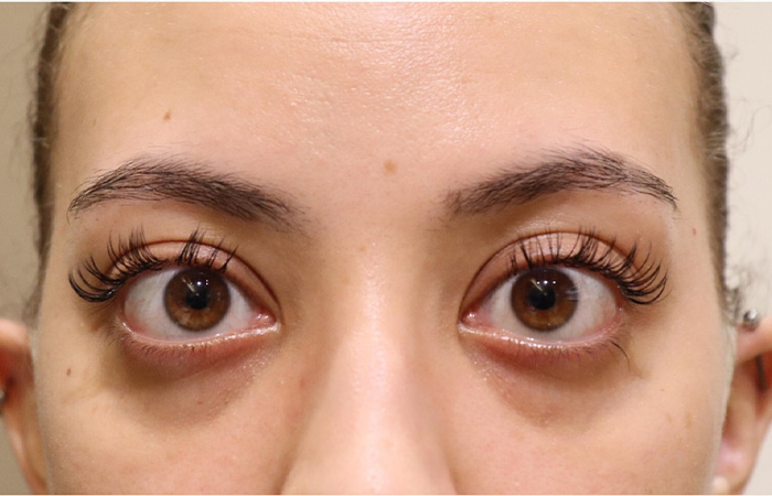
Pre
The eyes are large and give the impression that they stand out too much. The area of the whites of the eyes is large, making them look like Sanpaku eyes. It also looks like the eyes are a little far apart.
Post
This is a photo taken the day after surgery. You can see that the eyes are sunken and the area of the whites of the eyes has decreased. The distance between my eyes has become closer and my balance has improved. The black lower eyelid is due to internal bleeding.
Easy to understand and enlarge
Pre
I have enlarged the photo for clarity. The area of the white of the eye is large, and there is also the white of the eye between the cornea and lower eyelid.
Post
This is a photo taken the day after surgery. You can see that the upper eyelids are sunken and the area of the whites of the eyes is reduced. The white of the eye between the lower eyelid and cornea is gone, and the tripaku has resolved. The black lower eyelid is due to internal bleeding.
Next, let’s take a look at the photo from an angle.
Pre
It is noticeable that the eyes are very large. I think people with small eyes would be envious of them, but having them too big can also be a problem.
Post
In the photo taken the day after surgery, the area of the whites of the eyes has decreased and the impression that the eyes are large no longer appears. It has changed to a calmer impression.
Enlarge the photo of the right eye
Again, we may remove fat as much as possible if patient demands, but it may result worsening of impression.
Pre
The position of the eyebrows and the position of the eyeballs are approximately the same height.
Post
It’s the day after surgery. As the eyeballs become sunken, the upper eyelids become sunken, and as a result, the area of the whites of the eyes becomes smaller.
Case4.40 years old male
Pre
The eyes are bulging and the upper eyelids are bulging.
Post
His eyes became sunken and small. The swelling of the upper eyelids was reduced.
Conclusion
As a conclusion, any exophthalmos can be treated by orbital fat decompression surgery to some extent.
Risks associated with this treatment and their incidence
Internal bleeding may occur at the wound site due to surgery. At first, internal bleeding looks like a red bruise, but it turns yellow and moves downward under the skin due to gravity, disappearing in about 3 weeks.
The swelling will mostly subside within the first two weeks.
It takes about six months for complete disappearance. If a hematoma forms in the wound, surgery is required to remove it.
If there is no diplopia before surgery, eye movement disorders will appear after surgery, resulting in double vision. It often disappears by the next day, but it may remain.
If diplopia remains, it will gradually improve over 3 to 6 months, but diplopia remains in 0-3% of cases with lipectomy alone, 3-6% with lateral walls, and 10-65% with medial walls. It is said that then. In that case, strabismus surgery may be necessary. Even if double vision does not appear in frontal vision, there is a 10% chance that double vision will remain in peripheral vision (up, down, left, and right).
After the surgery, your pupils may dilate or it may become difficult to see up close, but this will gradually improve over about six months.
If damage occurs to the important nerves related to the eye or the blood vessels that feed them, vision and visual field problems may occur, leading to blindness. The incidence is approximately 1%, including mild cases.
Since the surgery is performed close to the brain, infection can lead to serious conditions.
The scar will gradually become less noticeable after surgery, but it may become noticeable or become a keloid. If the wound disintegrates after surgery, suturing will be required again. Orbital cellulitis may occur due to infection.