Orbital decompression, which removes the fat and bone behind the eyes for patients with bulging eyes,
It can also be performed for congenital exophthalmos that are not caused by Graves’ disease.
Proptophthalmia that is not due to Graves’ disease
Orbital decompression can also be performed for congenital proptosis that is not caused by Graves’ disease. People who are born with proptophthalmia, which makes their eyes appear as if they are protruding, do not have protruding eyes, but simply because the bones around their eyes have not developed properly. Very likely.
If tests for Graves' disease show no abnormalities, the patient will be diagnosed with congenital exophthalmia.
At our hospital, we perform a surgery called orbital decompression, which removes the fat and bone behind the eyes, for people with bulging eyes due to Graves' disease.We can apply this technique to treat people with bulging eyes who were born with bulging eyes. We also perform surgery.
However, health insurance cannot cover congenital bulging eyes, so you will have to pay for the entire cost out of pocket. However, if your eyes are too big, there is a good chance that you can make your eyes look more charming by making them smaller and modest. is.
About surgery for Graves’ disease and thyroid eye disease
Orbital fat decompression is less invasive to patients
Same-day surgery
both eyes at the same time
Remove only the increased fat
surgery without cutting the skin
In Graves’ disease and thyroid eye disease, the fat behind the eyeballs increases and the muscles that move the eyeballs swell, causing the eyeballs to move forward and causing a significant change in facial appearance.
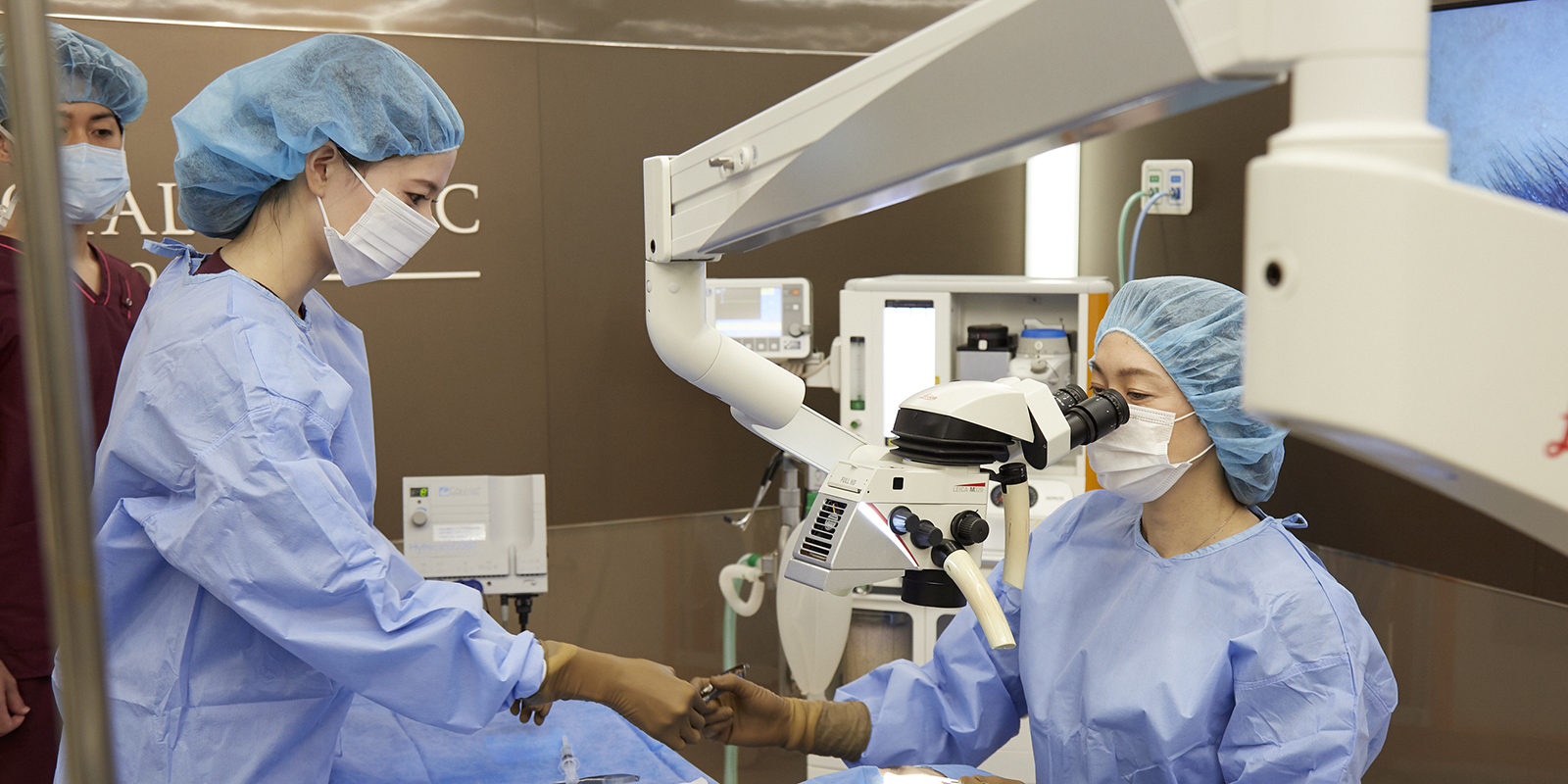
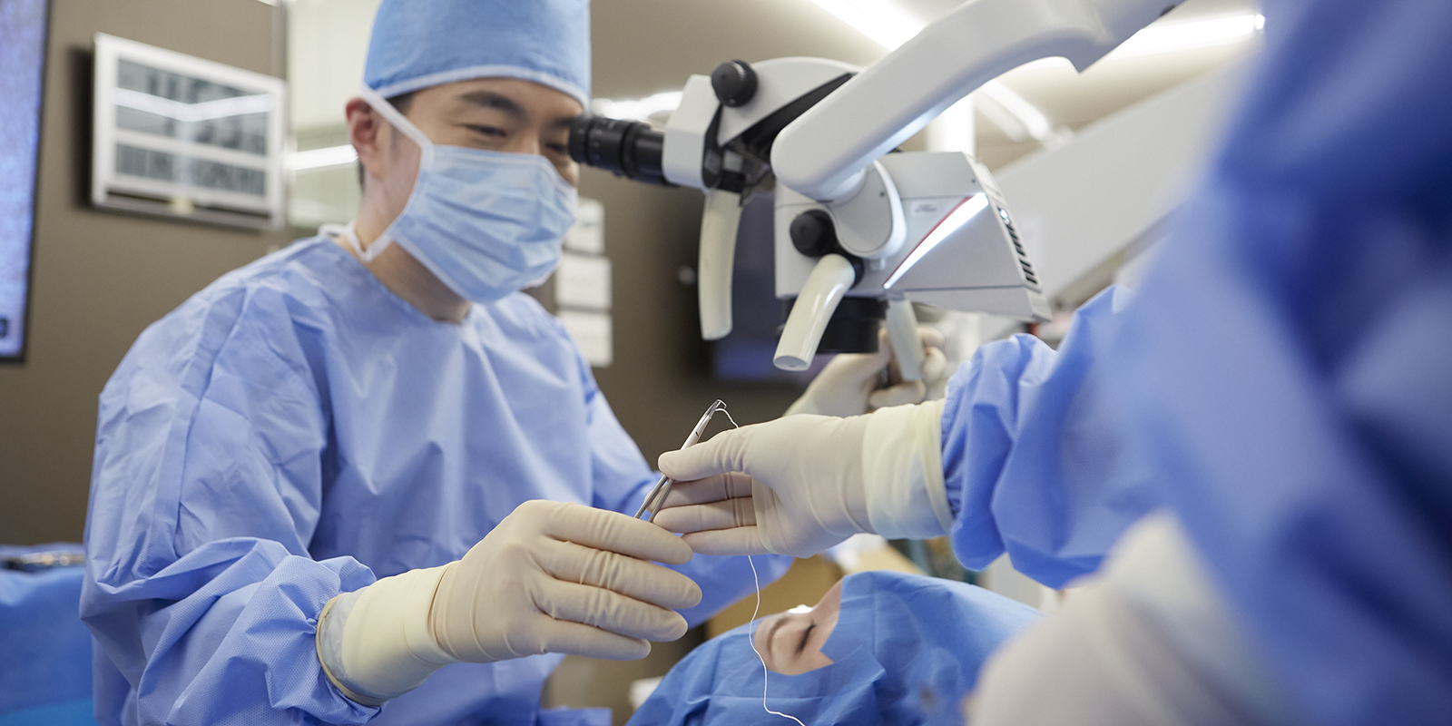
The orbital fat decompression surgery that we perform at our hospital is based on the cutting-edge medical care that I gained while studying abroad in the United States, and by gaining knowledge from famous overseas doctors and using a microscope to perform it in a more detailed manner in a Japanese style. This is a surgery that is the culmination of world knowledge that reduces tissue damage. There is no medical institution in the world that performs this surgery.
Same-day surgery is possible
This surgery avoids nerves and blood vessels related to visual function and only removes the increased fat, and requires extremely delicate and special techniques. Decompression surgery, which removes bones, was devised because there was no technology to remove fat while avoiding nerves and blood vessels, but it is physically taxing because it removes bones, and it may be difficult to do so due to post-operative pain and bleeding. Requires hospitalization for 2 weeks.
With orbital fat decompression at our clinic, both eyes are treated at the same time under general anesthesia, and you can go home the same day, minimizing the impact on your work and home life. Unfortunately, as it is a cutting-edge surgery, it is not covered by health insurance, but if you are suffering from an illness, we hope that you will choose orbital fat decompression at our hospital.
Graves’ disease and thyroid ophthalmopathy will be explained using case photos.
The face will look very different before and after treatment
See photos comparing before and after treatment
This woman had undergone the following treatments since the onset of Graves’ eye disease.
・Steroid pulse
・Orbital (cancer) decompression
・Additional fat removal
・Strabismus surgery (at another hospital)
If you compare before and after treatment, you will notice that your face is much prettier and your face looks completely different.
Next is a photo from the side
After surgery, the difference between before and after treatment was immediately obvious, and I was able to return to the beautiful facial features I had before the onset of the disease. It seems that Graves’ eye disease was once called a beauty disease, but looking at this case, it becomes clear that the idea that “one who develops the condition becomes a beautiful woman” is misguided.
What exactly is Graves’ disease?
The big problem with Graves’ disease and thyroid ophthalmopathy is that even if you receive intravenous or oral treatment and enter a stable period, once you recover like a cold, you will not be back to normal, and your appearance will be completely different from before. That’s it.
The problem is that most of the people who experience changes in appearance are young women. In English, this ugly bulging eyeball is called Disfiguring Proptosis.
It is sometimes incorrectly said that “protrusion of the eyeballs will go away once the internal medical values stabilize”, but once the exophthalmos protrudes, it will not return to its original state.
Can Graves’ disease/thyroid eye disease be cured?
Many people suffer from this condition, but there are only a few facilities nationwide that provide surgical treatment for Graves’ disease and thyroid eye disease.
Additionally, some ophthalmology facilities require hospitalization for several weeks as the surgery is quite extensive. For this reason, there are very few facilities that perform surgery for relatively mild proptosis.
For these people, we perform surgeries that emphasize cosmetic aspects as much as possible based on our specialized knowledge and experience. There are many patients who have been refused surgery at other hospitals because they are not suitable for surgery, but who undergo surgery at our facility. In addition, patients who have given up on the condition because it cannot be treated come back for surgery even though it has been a long time, even decades, since the onset of the disease. I hope you can consult with me even just once.
Surgery example of orbital decompression
Explaining orbital decompression using case photos
Compare photos before and after treatment
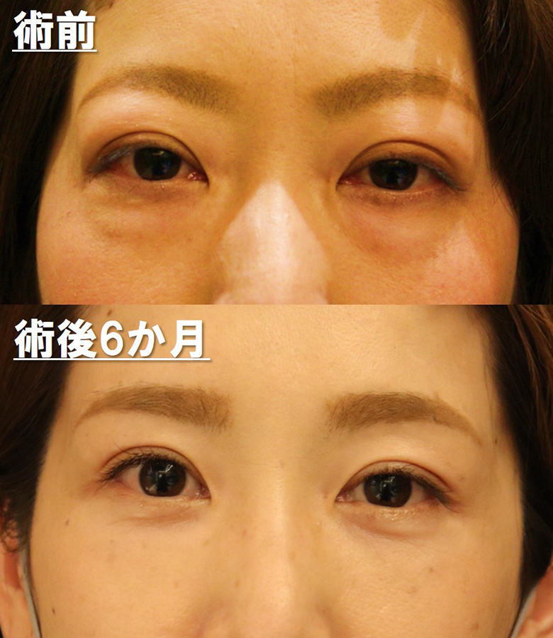
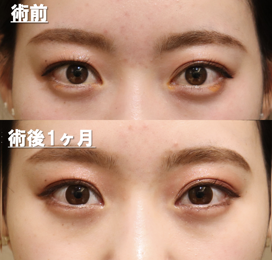
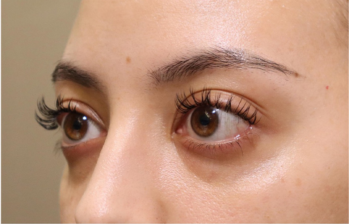
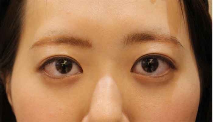
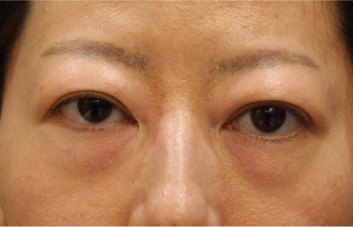
Please take a look at the pre-surgery photos. She has beautiful eyes with double eyelids, but the whites of her eyes are exposed a lot, and her upper eyelids look a little swollen, so she came to our clinic hoping to have these things corrected.
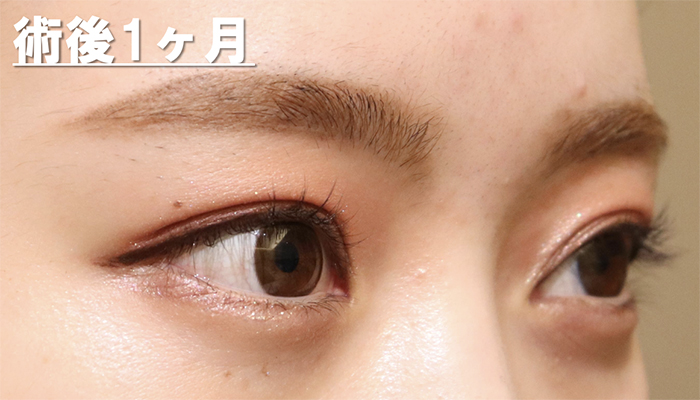
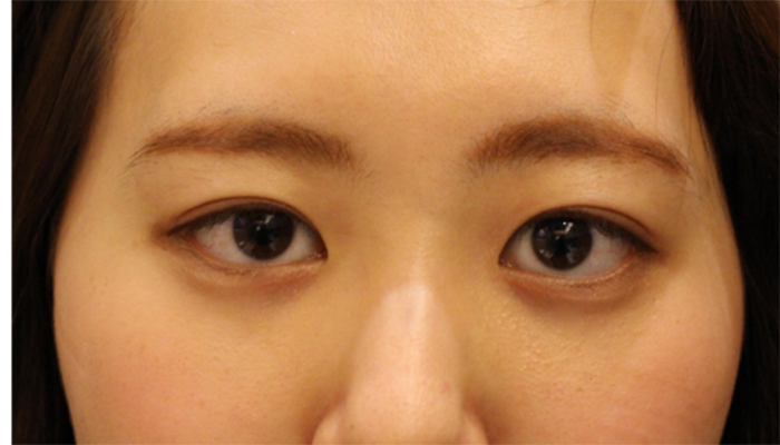
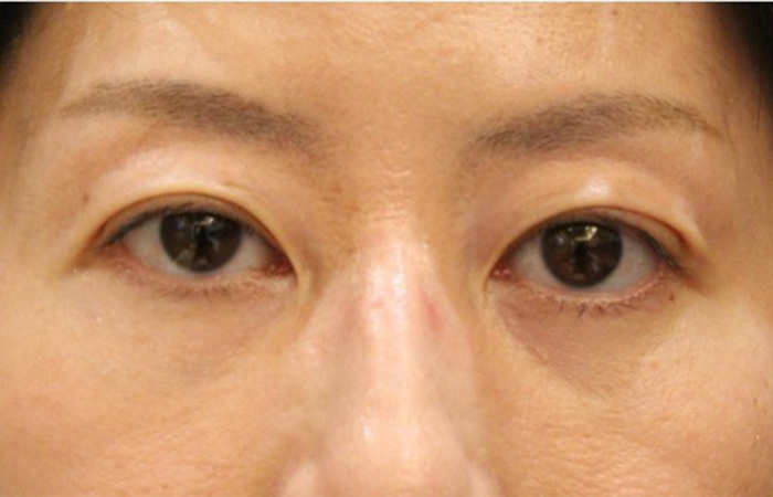
Next, please take a look at the photos taken 1 month after surgery.
There are still traces of bleeding on the lower eyelids and whites of the eyes. It usually takes 3-4 weeks for the bleeding to subside. You can see that the sunken eyes give a gentler impression.
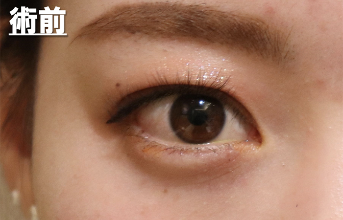
Easy to understand and enlarge
Pre
There is a bulge in the upper eyelids, giving the impression of being a little chubby. The area of the whites of the eyes is also large.
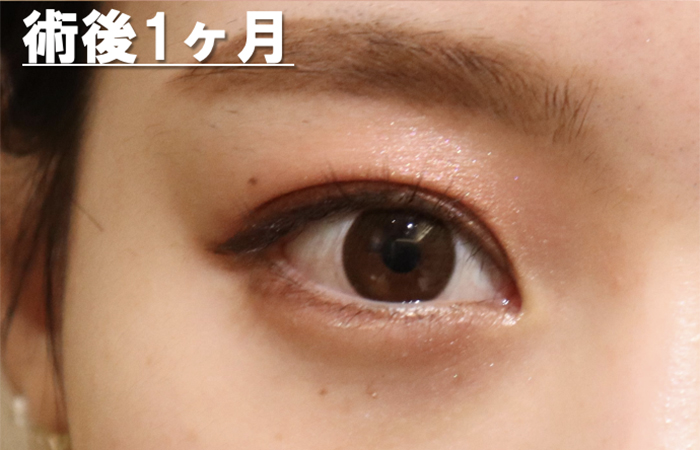
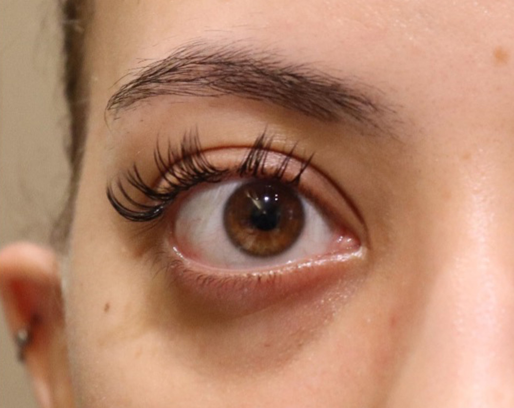
Post
It’s been 2 weeks since the fat was removed from the lower eyelid and behind the eyeball. I still have bleeding in the whites of my eyes and lower eyelids. The area of the whites of the eyes has decreased. Also, because the eyes are sunken, both the upper and lower eyelids cover the eyes, reducing the exposure of brown eyes.
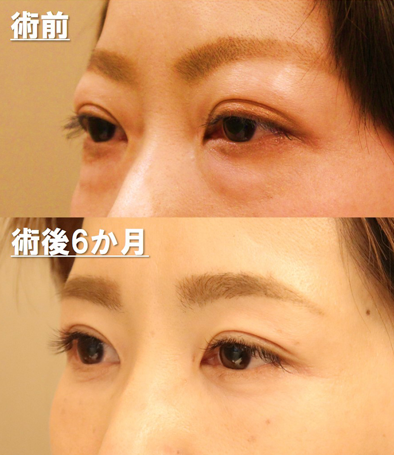
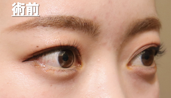
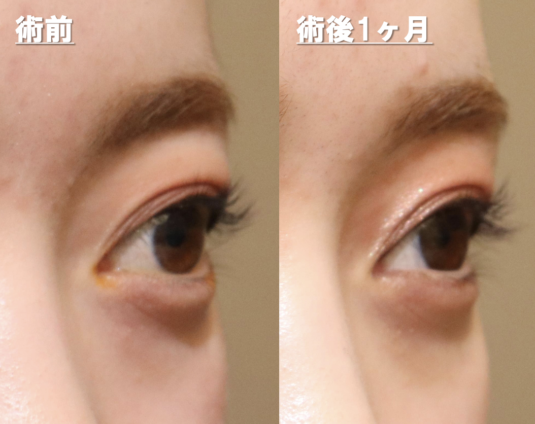
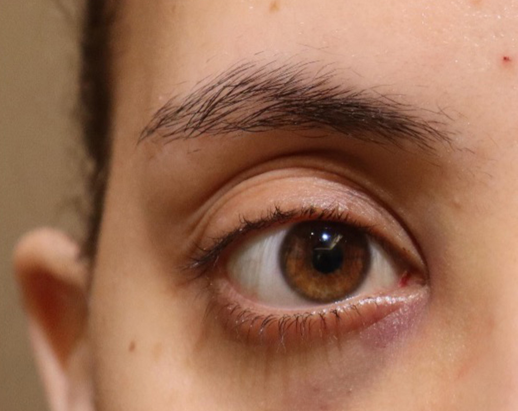
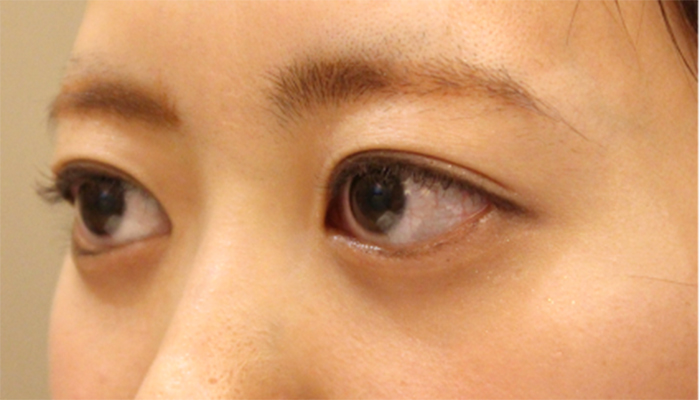
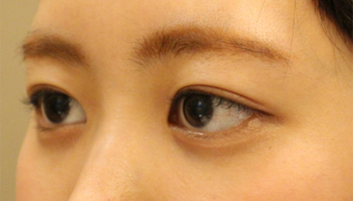
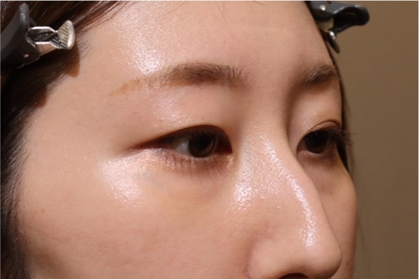
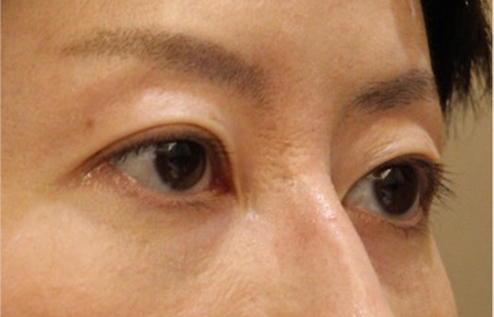
Next is a photo taken from an angle.
Pre
She has beautiful eyes with double eyelids, but the whites of her eyes are exposed a lot, and her upper eyelids have a slightly swollen shape.
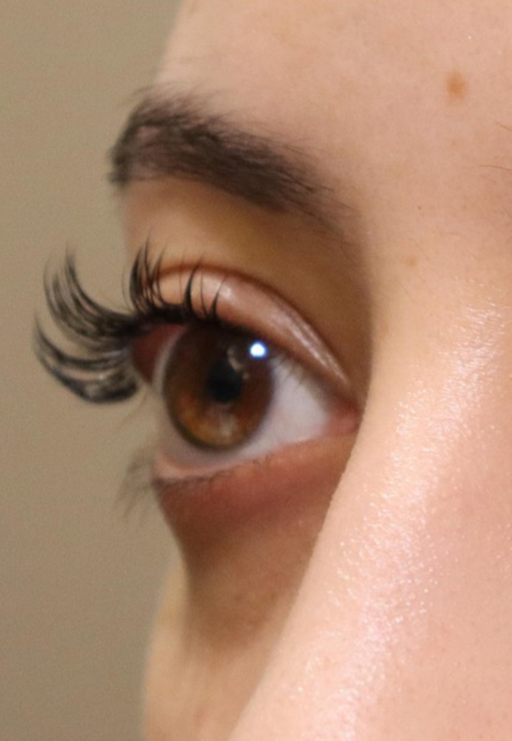
Post
Comparing the area of the whites of the eyes, the area of the whites of the eyes is smaller after surgery. Her eyes are a little smaller, giving her a more beautiful look. My upper eyelids also feel refreshed.
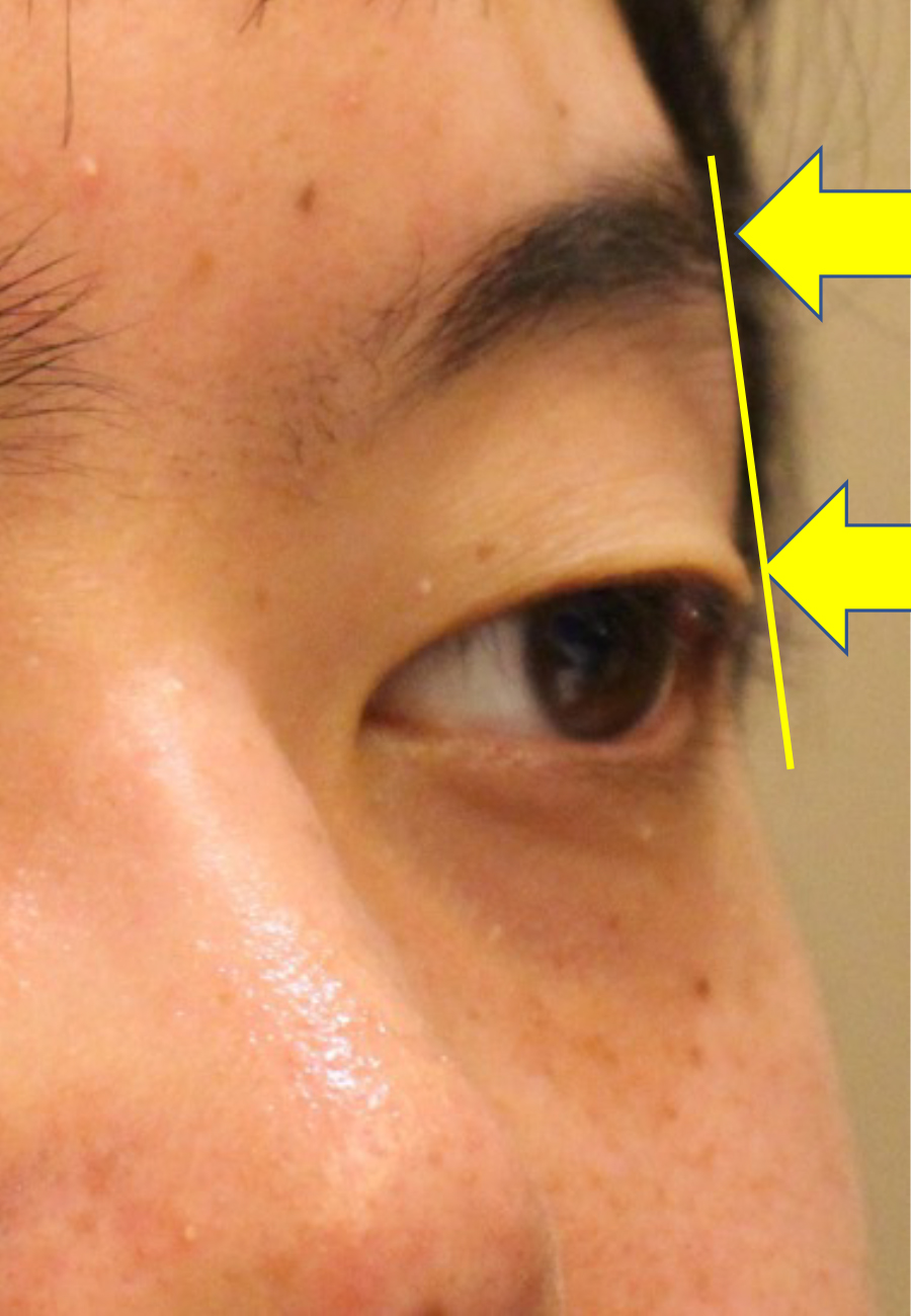
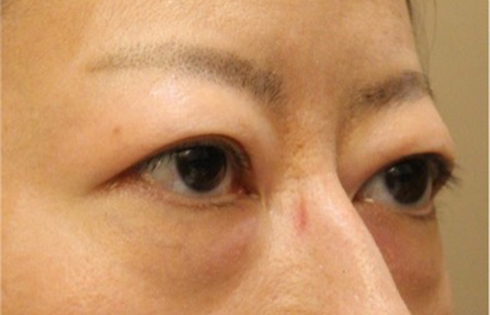
Please pay attention to your left eye
Compare photos before and after treatment
Comparing the left upper eyelid in an oblique photo, you can see that the puffiness has disappeared and the upper eyelid, which used to protrude more than the eyebrows, has become more depressed than the eyebrows after surgery.
In this way, you are born with large eyes (protruding eyes), and our clinic also performs corrections for this. If you have a similar problem, please feel free to contact us.
Risk
Risk of complications
Bleeding, infection, double vision, visual field disturbance, visual impairment, general anesthesia
Cases of congenital bulging eyes
Explaining a case of congenital exophthalmia
Deepening of orbital sulcus cause completely different appearance which may bring impression of aged person.
Compare photos before and after treatment
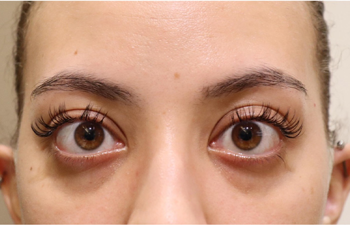
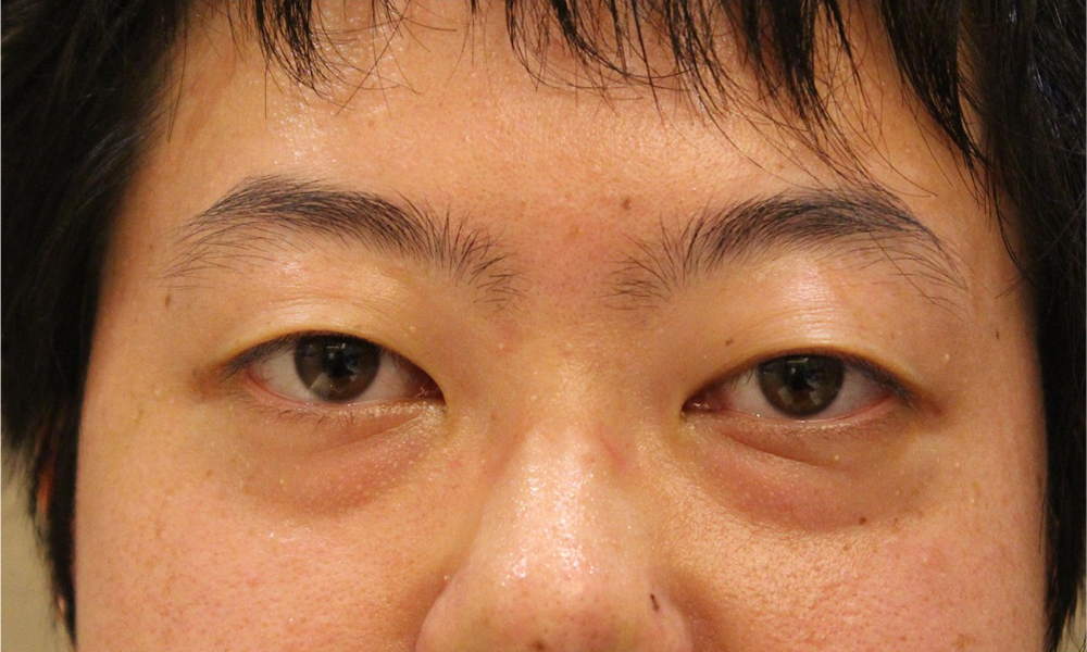
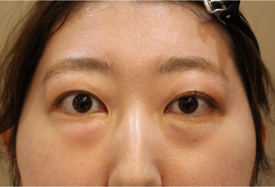
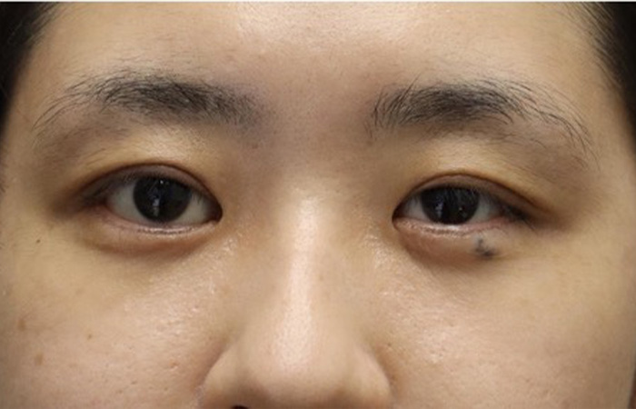
Pre
The eyes are large and give the impression that they stand out too much. The area of the whites of the eyes is large, making them look like Sanpaku eyes. It also looks like the eyes are a little far apart.
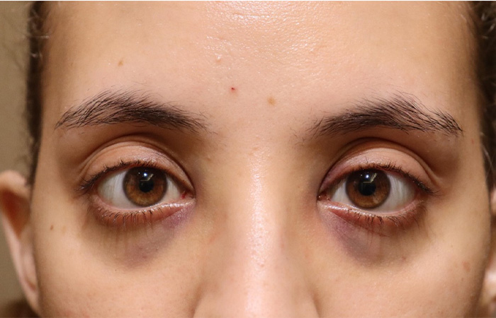
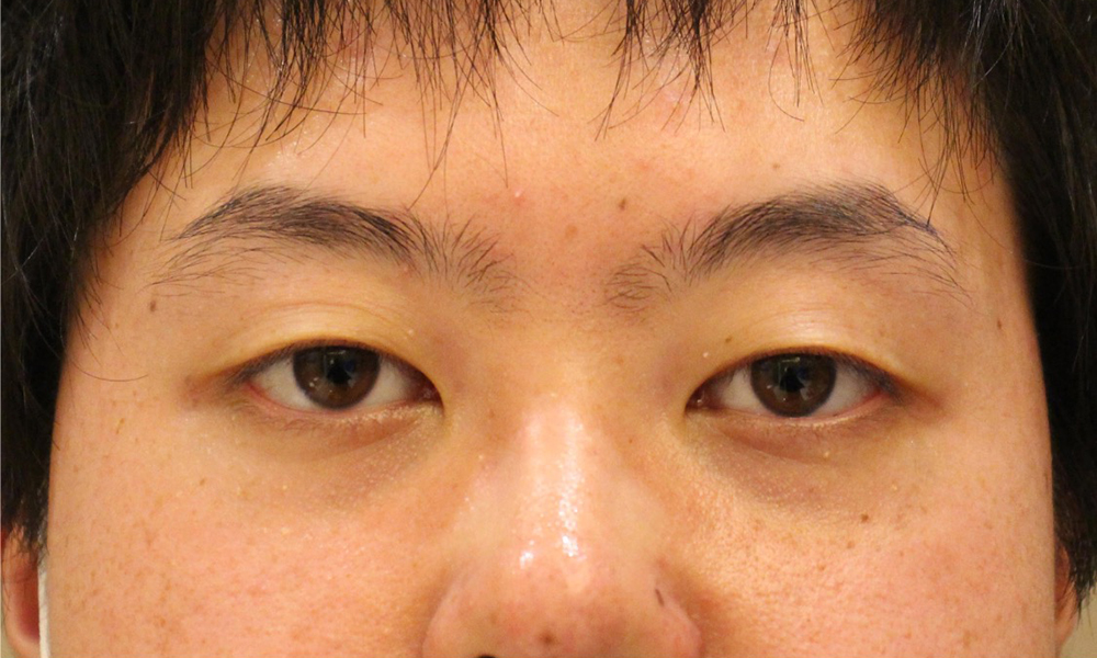
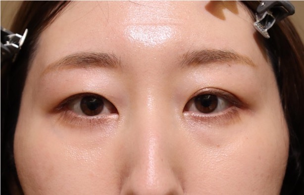
Post
This is a photo taken the day after surgery. You can see that the eyes are sunken and the area of the whites of the eyes has decreased. The distance between my eyes has become closer and my balance has improved. The black lower eyelid is due to internal bleeding.
Easy to understand and enlarge
Pre
I have enlarged the photo for clarity. The area of the white of the eye is large, and there is also the white of the eye between the cornea and lower eyelid.
Post
This is a photo taken the day after surgery. You can see that the upper eyelids are sunken and the area of the whites of the eyes is reduced. The white of the eye between the lower eyelid and cornea is gone, and the tripaku has resolved. The black lower eyelid is due to internal bleeding.
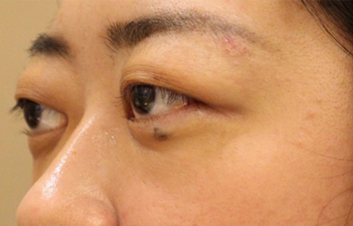
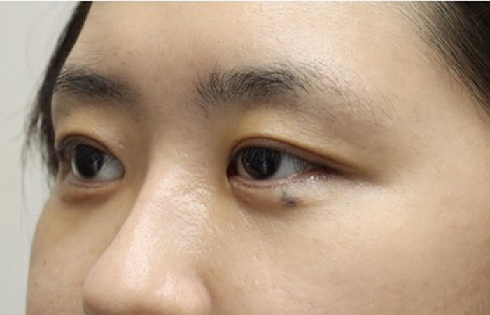
Next, let’s take a look at the photo from an angle.
Pre
It is noticeable that the eyes are very large. I think people with small eyes would be envious of them, but having them too big can also be a problem.
Post
In the photo taken the day after surgery, the area of the whites of the eyes has decreased and the impression that the eyes are large no longer appears. It has changed to a calmer impression.
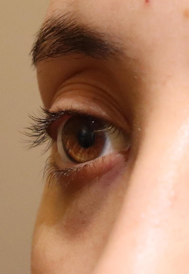
Enlarge the photo of the right eye
Again, we may remove fat as much as possible if patient demands, but it may result worsening of impression.
Pre
The position of the eyebrows and the position of the eyeballs are approximately the same height.
Post
It’s the day after surgery. As the eyeballs become sunken, the upper eyelids become sunken, and as a result, the area of the whites of the eyes becomes smaller.
Other cases of orbital decompression surgery
Case1 Approximately 20 years old female
Pre
A condition in which the eyes protrude greatly and the area of the whites of the eyes is large.
Post
The eyes have become smaller and the area of the whites of the eyes has also become smaller.
Pre
A condition in which the eyes protrude greatly and the area of the whites of the eyes is large.
Post
Since the eyes are smaller, the redness in the whites of the eyes has also been reduced.
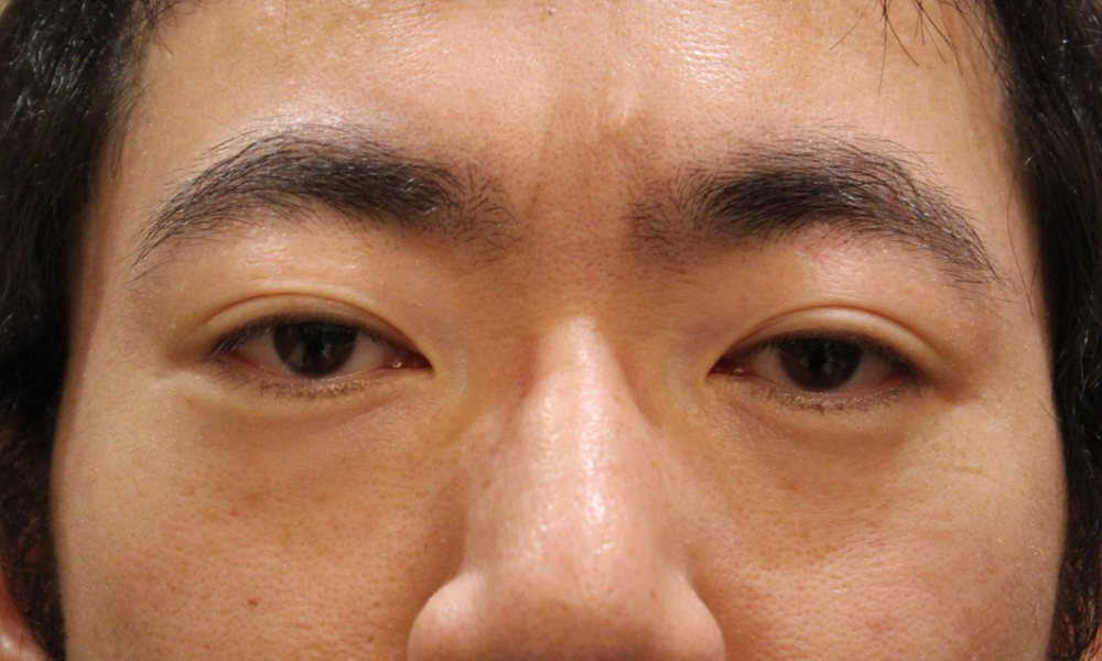
Case2 30 years old Male
Orbital fat decompression can treat congenitally proptotic patient.
Pre
Her eyes bulge out, her upper eyelids bulge, and her lower eyelids have dark circles.
Post
Eyes are smaller, upper eyelid bulge and lower eyelid dark circles have improved.
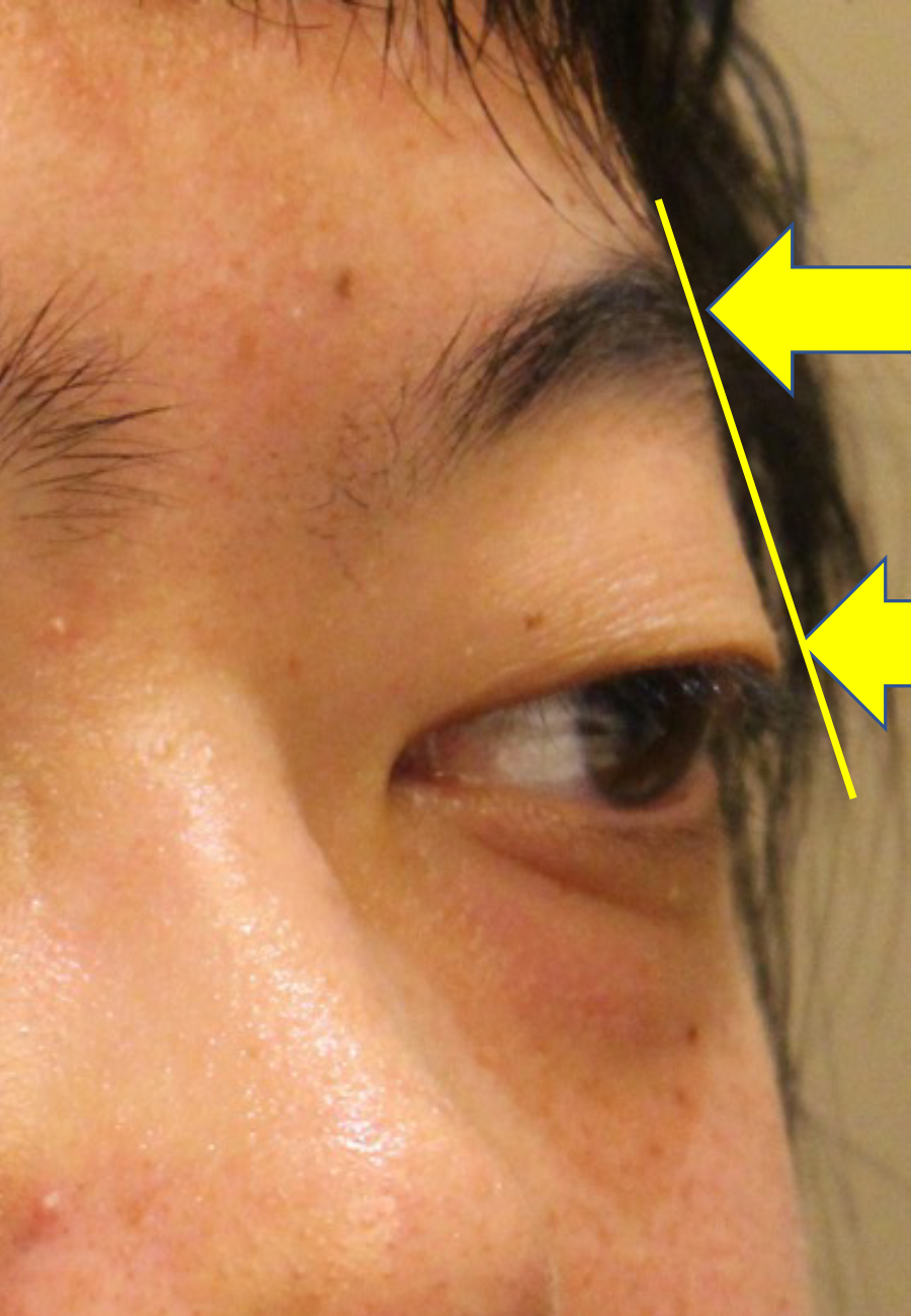
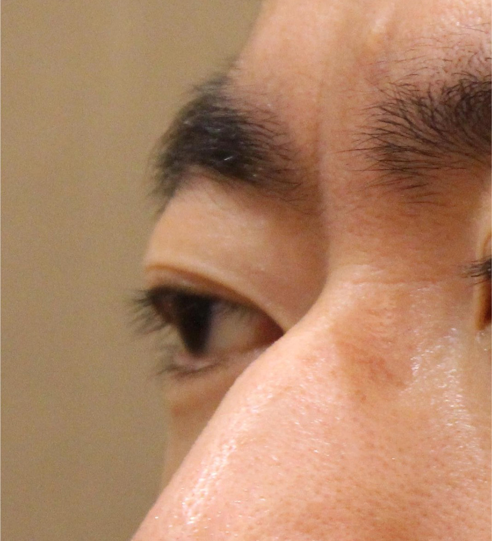
However, from the side, you can understand the change.
Comparing the top of eyelid and eyebrow, it is remarkable eyeball position changed
Different from thyroid eye disease, congenital proptosis do not have increasing volume of fat.
So there is limitation to remove the fat.
Pre
The upper eyelids are swollen and the lower eyelids have dark circles.
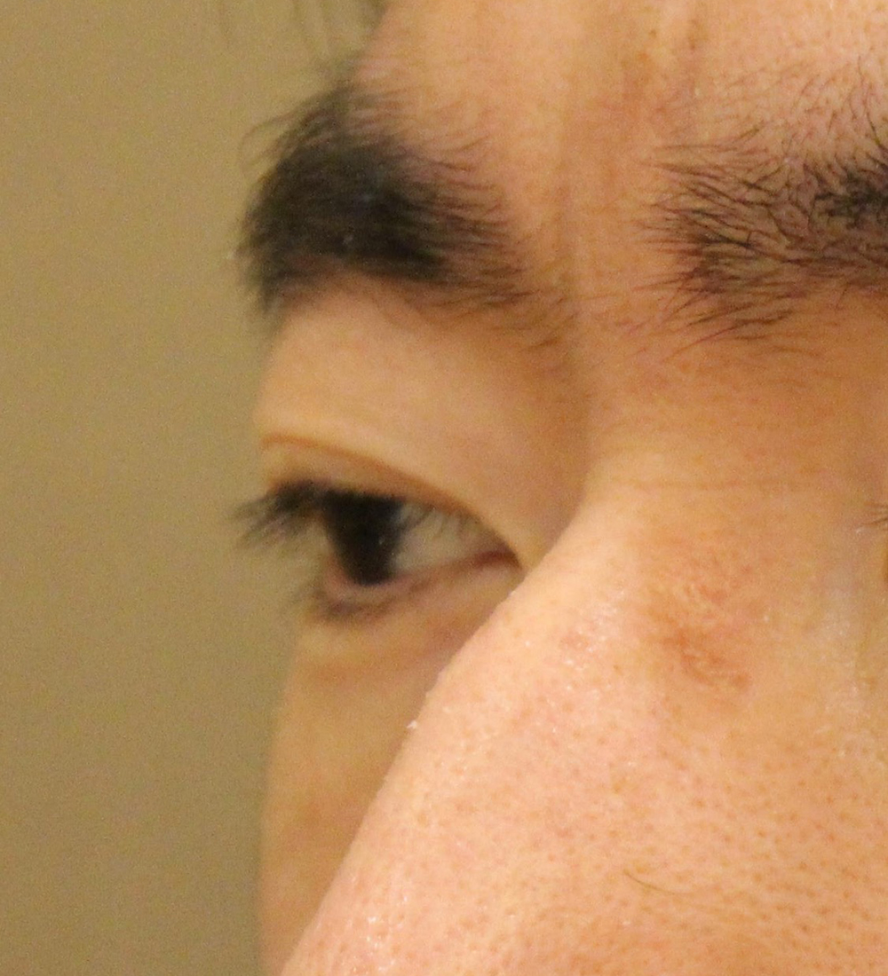
Post
Eyes are smaller, upper eyelid bulge and lower eyelid dark circles have improved.
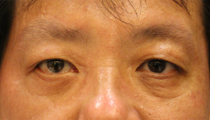
Case3 Approximately 40 years old male
Pre
The eyes are bulging and the upper eyelids are bulging.
Post
His eyes became sunken and small. The swelling of the upper eyelids was reduced.
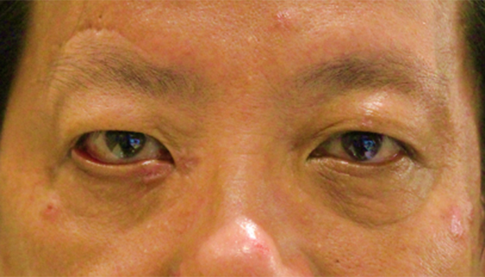
Case 4 Approximately 50-year-old male (after multiple surgeries for right facial nerve paralysis at another hospital)
Pre
The eyes are bulging and the upper eyelids are bulging. There is a clear surgical scar above the right eyebrow. Although multiple surgeries including fascia transplantation were performed on the right lower eyelid, ectropion remains.
Post
His eyes became sunken and small. The swelling of the upper eyelids was reduced. The right lower eyelid ectropion has improved and the left-right difference in eye shape has disappeared.
Case5 30 years old Female
Pre
She has fatty hyperplasia of both upper and lower eyelids with ocular protrusion.
Post
Both upper and lower eyelids show improvement in adipogenesis.There seem to be no fat gain due to thyroid eye disease.
Case6 40 years old Female
This is the result after fat decompression. We performed both upper and lower fat decompression for this case.
Pre
Before surgery, upper and lower eyelid was puffy and swollen.
Post
After surgery, the eyelid become thin, skinny.Especially, please check far side of upper eyelid, eyelid become sharp and natural.
Case7 30 years old Female
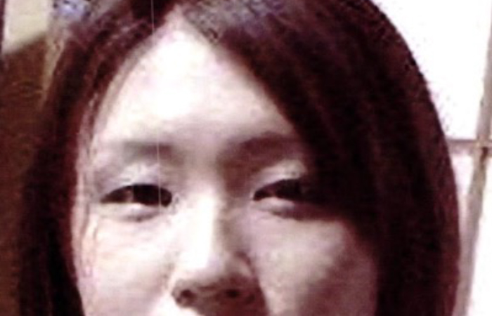
Pre
After onset of thyroid eye disease, her appearance has completely changed.
Post
After two types of fat decompression, she was back to her old self.Thyroid eye disease make both eyes apart, Removing fat of just behind the lacrimal caruncle, where is medial position of eyeball, eyes become closer.
Risks associated with this treatment and their incidence
Internal bleeding may occur at the wound site due to surgery. At first, internal bleeding looks like a red bruise, but it turns yellow and moves downward under the skin due to gravity, disappearing in about 3 weeks.
The swelling will mostly subside within the first two weeks.
It takes about six months for complete disappearance. If a hematoma forms in the wound, surgery is required to remove it.
If there is no diplopia before surgery, eye movement disorders will appear after surgery, resulting in double vision. It often disappears by the next day, but it may remain.
If diplopia remains, it will gradually improve over 3 to 6 months, but diplopia remains in 0-3% of cases with lipectomy alone, 3-6% with lateral walls, and 10-65% with medial walls. It is said that then. In that case, strabismus surgery may be necessary. Even if double vision does not appear in frontal vision, there is a 10% chance that double vision will remain in peripheral vision (up, down, left, and right).
After the surgery, your pupils may dilate or it may become difficult to see up close, but this will gradually improve over about six months.
If damage occurs to the important nerves related to the eye or the blood vessels that feed them, vision and visual field problems may occur, leading to blindness. The incidence is approximately 1%, including mild cases.
Since the surgery is performed close to the brain, infection can lead to serious conditions.
The scar will gradually become less noticeable after surgery, but it may become noticeable or become a keloid. If the wound disintegrates after surgery, suturing will be required again. Orbital cellulitis may occur due to infection.
Author information
Tomoyuki Kashima
/ MD, PhD
Over the past 10 years, we have performed more than 10,000 eye plastic surgeries in Japan and the United States. I aim to provide the cutting-edge medical knowledge and experience I have gained to patients in need in Japan.