Our hospital strives to perform surgeries that minimize the burden on patients, such as post-operative pain and scars, more than any other medical institution in Japan. Please do not hesitate to contact us.
ContactOrbital tumor
目次
- 1 Surgery to remove orbital tumors with as little scarring as possible
- 2 Orbital tumor
- 2.1 Approximately 70 years old Female Right huge intraconal orbital tumor Operation refused by neurosurgeon
- 2.2 Explanatory video of myocone intraorbital tumor removalOcculofacial Clinic Tokyo Dr. Tomotaka Kashima
- 2.3 Case 2. Diagnosed with orbital tumor, but A male patient in his 50s came to our hospital after being told that surgery was not possible.
- 2.4 Explanatory video of myocone intraorbital tumor removalOculo facial Clinic Tokyo Director Tomotaka Kashima
- 2.5 Approximately 50 years old Female Left extraconal orbital tumor
- 2.6 Approximately 70 years old Male Right lacrimal grand tumor
- 2.7 Please feel free to contact us first.
Surgery to remove orbital tumors
with as little scarring as possible
There are only a few medical institutions specializing in orbital tumors in Japan.
There are only a few medical institutions in Japan that specialize in orbital tumors. Moreover, even if the medical institution is famous for treating orbital tumors, there are almost no medical institutions in Japan that specialize in the surgery called orbital tumor removal.
Therefore, there are many patients (and doctors) who are hesitant to undergo surgery because they are told that the tumor cannot be removed unless the head is opened (craniotomy).
Because of our extensive treatment history, we are able to perform surgery without leaving any scars.
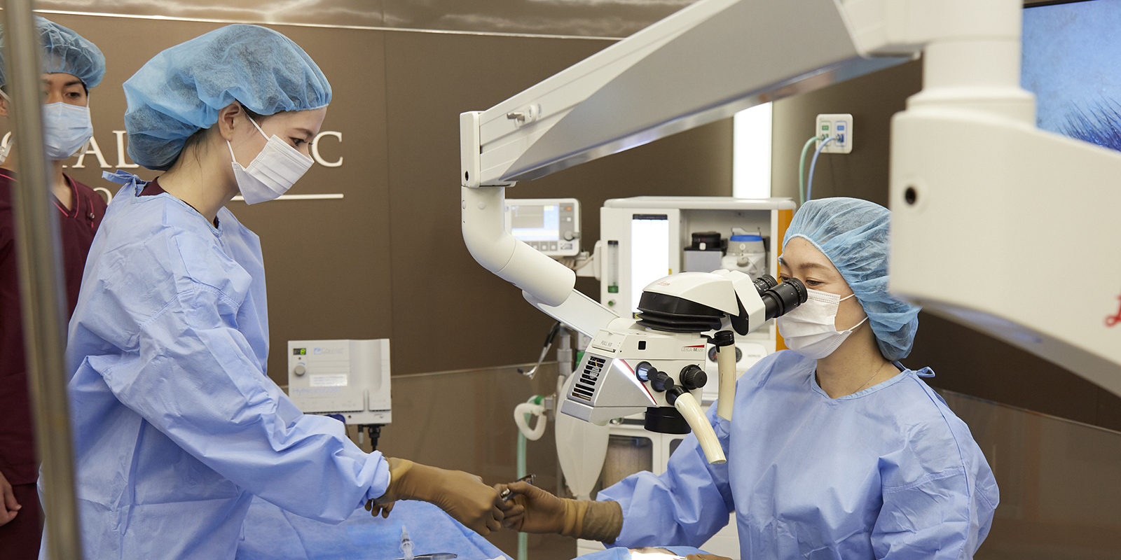
We perform many orbital surgeries every day, including orbital decompression for thyroid eye disease and orbital blowout fractures.
Utilizing that experience, we have performed many surgeries to remove orbital tumors that were considered difficult at other hospitals.
The incision site is not a traditional large incision, but rather a location that leaves as few scars as possible and allows direct access to the tumor.
We aim to perform surgeries that are as gentle to the patient as possible and are gentle on the eyeballs. If you have any problems, please feel free to contact us.
Orbital tumor
Approximately 70 years old Female
Right huge intraconal orbital tumor
Operation refused by neurosurgeon


This patient is approximately 70-year-old woman. She had been seeing a neurosurgeon, but because there’s a risk of blindness, she has not been able to undergo surgery until now. Gradually, the protrusion of right began to interfere with daily life, not only in terms of function but also in terms of appearance. She came to our hospital looking for a medical institution that could treat her right eye
Intraconal tumor bigger than eyeball

MRI shows a tumor larger than the eye in the posterior part of the eye; both T1 and T2 have similar signal intensity as the vitreous cavity. The border is clear. The diameter of the tumor is about 35 mm. How would you remove this tumor?
I chose to approach through a lateral canthotomy incision.
I started from lateral canthotomy. Cut into until periosteum, and then removed lateral wall of orbit.
The key of orbital tumor removal is how to use the malleable retractors.
Using 2 malleable retractors, separate the tumor from orbital tissue, and then, remove it.
3 month post-operation


The tumor was successfully removed and vision was restored.
Because of the very large tumor, the eye is rather concave after removal.
Comparison of before and after surgery.

The upper row is preoperative and the lower row is postoperative. You can see that the huge tumor was removed and no scar remains on the skin. Compared to before, the protrusion of the eyeball has improved and the vision, which was almost blind, has recovered to almost normal. Obviously, her quality of life improved due to cosmetic change.
Surgery video
Explanatory video of myocone intraorbital tumor removal
Occulofacial Clinic Tokyo
Dr. Tomotaka Kashima
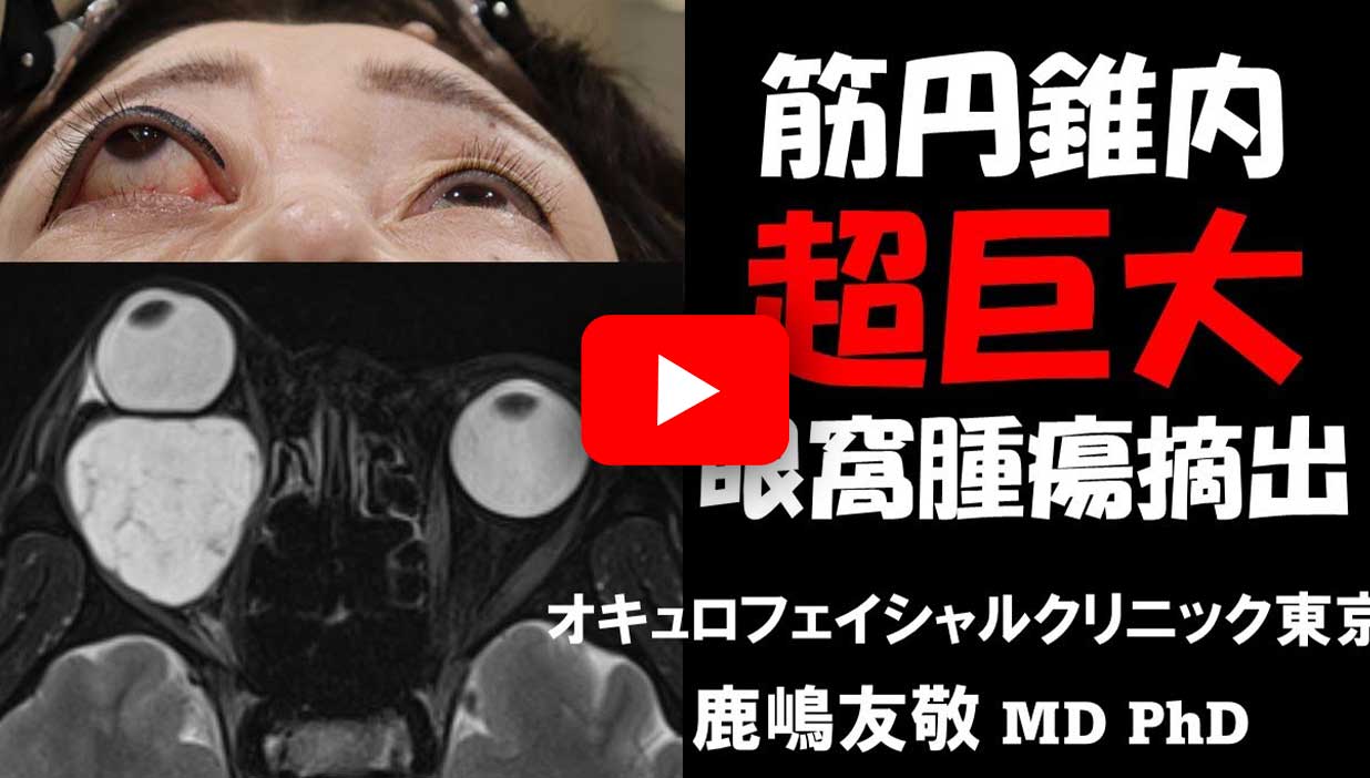
*This is an explanatory video of the actual surgery, so if you are not comfortable with surgical videos, please do not watch it.
Case 2. Diagnosed with orbital tumor, but A male patient in his 50s came to our hospital after being told that surgery was not possible.
Male, about 50 years old. Three years ago, I was diagnosed with an orbital tumor at a university hospital. Although he was told that surgery was not possible, he was referred to our hospital by a friend.
Preoperative case photo

There is exophthalmia in the right eye, and the eye appears swollen compared to the left eye.

The proptosis of the right eye is noticeable compared to the left eye.
Post-operative case photos

The proptosis of the right eye has improved. The scratches around the corners of the eyes are not noticeable either.

There is improvement in ocular protrusion of the right eye.
Preoperative MRI

It turns out that there is a tumor behind the eyeball, pushing it up.
Post-operative MRI

The tumor was removed cleanly and the eyeball was sunken.
Surgery video
Explanatory video of myocone intraorbital tumor removal
Oculo facial Clinic Tokyo
Director Tomotaka Kashima
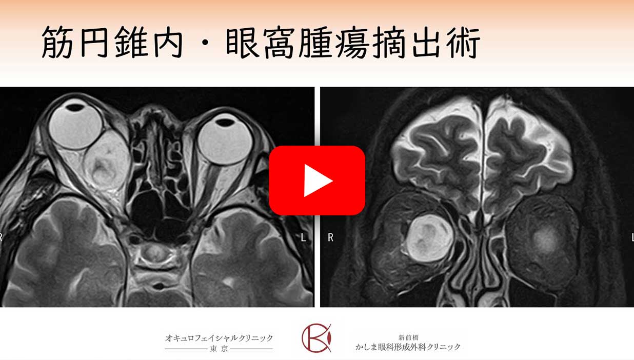
*This is an explanatory video of the actual surgery, so if you are not comfortable with surgical videos, please do not watch it.
Approximately 50 years old Female
Left extraconal orbital tumor


She is approximately 50 years old. A left orbital tumor is present below the eyeball. The increased size of the tumor has caused upward pressure on the eyeball, resulting in deviation.
This surgery was done with swinging eyelid approachIn Inferolateral quadrant, there is no important tissue, so I finished it in 18 mins. . I put this video on Youtube.

Postoperatively, differences in left and right eyelid shape and eye position have been resolved.
After surgery she regained symmetrical eyes cosmetically.
Approximately 70 years old Male
Right lacrimal grand tumor


The right eye is compressed by the tumor, causing not only ocular protrusion but also downward deviation of the eyeball.
Right Lacrimal gland tumor


MRI shows a tumor in the lacrimal gland area of the right orbit, compressing the eye.
I choose lid crease approach. Dissecting into the periosteum and expose bone.
And then, after bone removal, I removed lacrimal gland tumor.
Lacrimal gland tumor have loose adhesion laterally and superiorly, so you should start with that area.
It has tight adhesion inferiorly and medially, I recommend to loosen posterior side of lacrimal gland and dislocate it.


After tumor resection, ocular protrusion and downward deviation of the eyeball improved. But he developed and blepharoptosis.
Comparison of before and after surgery.

It can be seen that the preoperative protrusion of the eyeball has cosmetically improved. The tumor has stretched the levator aponeurosis, resulting In a blepharoptosis. This will be fixed by aponeurotic repair.
Please feel free to contact us first.