Indications:
This surgery is primarily indicated for individuals with extremely weak eyelid-lifting muscles (mainly congenital).
目次
Ptosis is referred to as the drooping of the eyelid. Normally, the position of the upper eyelid is 0.5 to 1.0 mm below the upper edge of the cornea. If the upper eyelid is lower than normal, it is considered to be ptosis. If ptosis is present at birth or within the first year of life, it is called congenital ptosis.
In most cases, congenital ptosis occurs alone and is not related to any systemic disease. There is no gender difference in congenital ptosis, and it can affect one or both eyes.
In many cases of congenital ptosis, there is no known cause, but in some cases, it occurs due to heredity (autosomal dominant). Histologically, it has been revealed that the levator muscle and aponeurosis tissues in patients with congenital ptosis are replaced by adipose and fibrous tissues.
Congenital ptosis exists from infancy, which can lead to a condition called "amblyopia" where vision does not develop. Amblyopia can occur due to visual impairment caused by the obstruction from ptosis, or it can result from astigmatism caused by corneal distortion, leading to visual impairment and consequently amblyopia.
Amblyopia can be an indication for early surgery. Also, the appearance in cases of ptosis is often not aesthetically pleasing, and surgery is recommended in such cases.
Not all patients with congenital ptosis require surgery, but it is necessary to carefully monitor children for the possibility of amblyopia.
Since amblyopia is less likely to be reversible after the age of 7 to 10, it is very important to perform surgery before this age to maintain vision.
Also, since vision tests become possible at the age of 3 to 4 years, diagnosis of amblyopia cannot be performed until after this period.
Therefore, it is considered appropriate to diagnose the presence or absence of amblyopia during the period from 3 to 7 years, and surgery should be performed if amblyopia is observed or suspected (except in cases of severe ocular torticollis).
Furthermore, ptosis can have a negative impact on development due to appearance issues.
In congenital ptosis, due to its physical, functional, and psychological impacts, surgery is recommended at some point, using one of various methods. The method of surgery varies depending on treatment goals, the underlying diagnosis, and the degree of levator function. While the main purpose of the surgery is functional, improving the appearance by making the eyelid shape and the width of the double eyelid symmetrical is also a goal.
Surgery for congenital ptosis can be performed at any age depending on the severity of the condition, but early surgery may be necessary, especially in cases of severe amblyopia or ocular torticollis. In severe cases of ocular torticollis, the extreme head-up posture can disrupt body balance and delay development during infancy and early childhood. If early surgery is not necessary, it is usually performed after the age of 3 or 4. Waiting until this age allows for more accurate measurements before surgery.
Regardless of the surgical technique used, it may become difficult to close the eyes, but this is generally not a problem until the 20s or 30s as the corneal surface is relatively resistant to dryness. Conversely, performing surgery in middle and older age can lead to corneal surface injuries, causing pain and foreign body sensation, and sometimes corrective surgery is required.

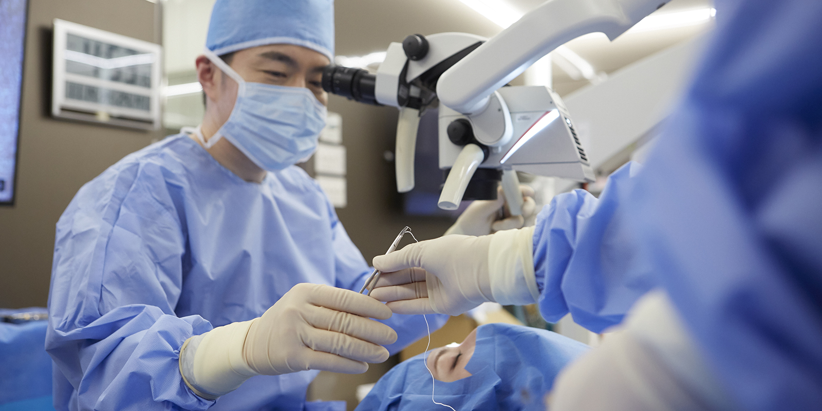
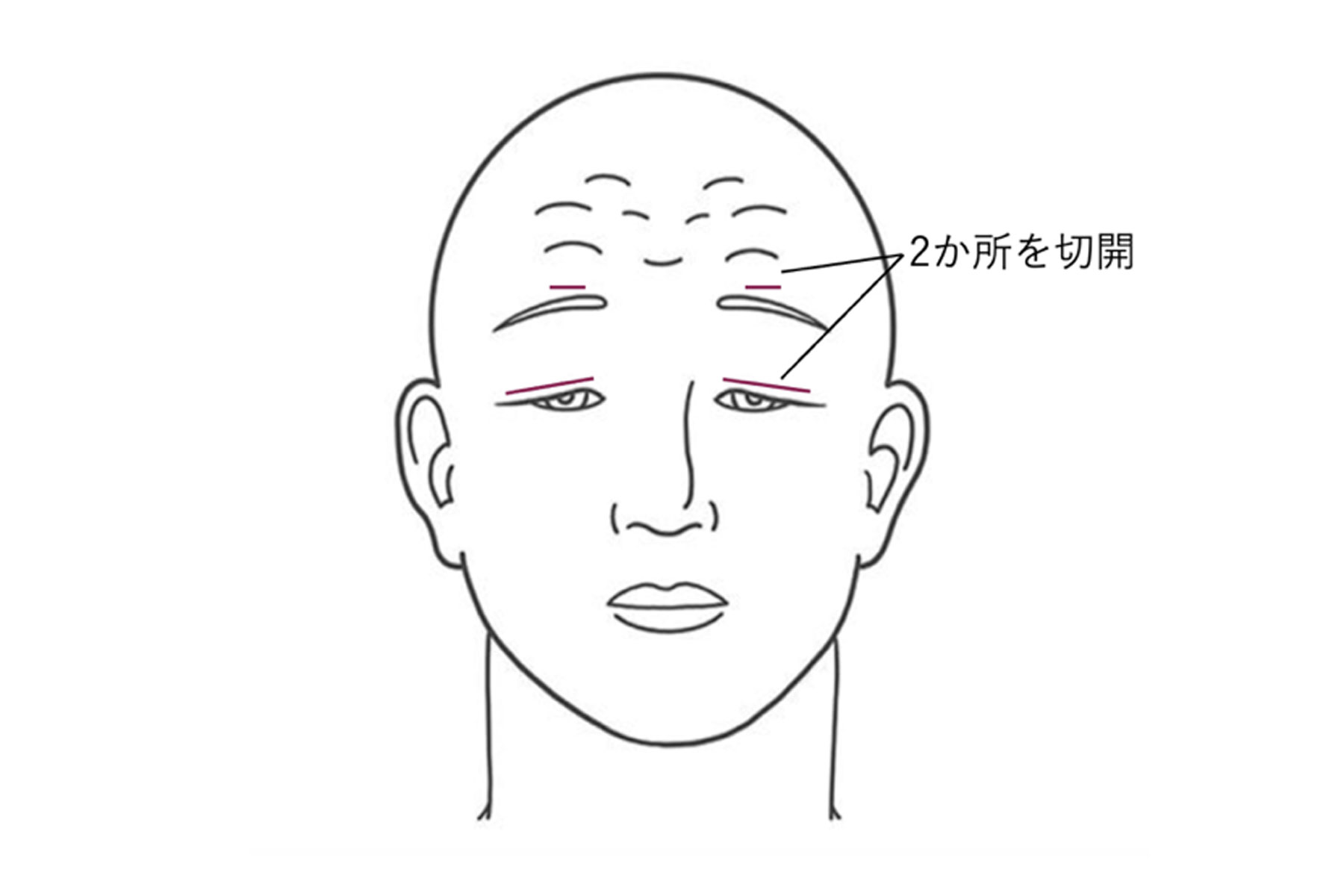
This surgery involves incising the double eyelid area to shorten the complex of the levator aponeurosis and Müller’s muscle. The skin incision is made so that it is hidden behind the original double eyelid or a newly created double eyelid.
To perform levator shortening, moderate levator function is necessary. If the levator function is more than 4mm but less than 6mm, a levator resection of more than 22mm is recommended. If the levator function is between 6mm and 8mm, the resection should be 16mm to 18mm. If levator function exceeds 8mm, a levator resection of 10mm to 13mm is required. If levator function is less than 4mm, or if there is an upward movement impairment, there may not only be insufficient eyelid elevation, but there is also a significant risk of severe corneal injury.
Individuals with a weak Bell's phenomenon (upward movement of the eyeball when the eyes are closed), decreased corneal sensitivity, or reduced tear production may be at risk of developing corneal abrasions.
Patients may be unable to close their eyelids during sleep for several weeks to months after surgery. Although the issue of eyelids remaining open during sleep improves over time, some degree of it may persist.
This surgery is primarily indicated for individuals with extremely weak eyelid-lifting muscles (mainly congenital).
This procedure connects the eyebrow to the eyelid, allowing the patient's eyelid to be lifted through the elevation of the eyebrow. In cases of severe unilateral congenital ptosis, the surgery may be performed bilaterally to achieve symmetry.
Materials used can include the patient’s own fascia lata, fascia lata from a tissue bank, non-absorbable suture materials (such as Prolene and nylon), silicone bands, silicone rods, and Gore-Tex sheets.
Individuals with a weak Bell's phenomenon (upward movement of the eyeball when the eyes are closed), decreased corneal sensitivity, or reduced tear production may be at risk of developing corneal abrasions.
Patients may be unable to close their eyelids during sleep for several weeks to months after surgery. Although the issue of eyelids remaining open during sleep improves over time, some degree of it may persist.
Surgical Method: This procedure connects the eyebrow to the eyelid, allowing the patient’s eyelid to be lifted through the elevation of the eyebrow. In cases of severe unilateral congenital ptosis, the surgery may be performed bilaterally to achieve symmetry. Materials used can include the patient’s own fascia lata, fascia lata from a tissue bank, non-absorbable suture materials (such as Prolene and nylon), silicone bands, silicone rods, and Gore-Tex sheets. Individuals with a weak Bell’s phenomenon (upward movement of the eyeball when the eyes are closed), decreased corneal sensitivity, or reduced tear production may be at risk of developing corneal abrasions. Surgical Outcomes: Patients may be unable to close their eyelids during sleep for several weeks to months after surgery. Although the issue of eyelids remaining open during sleep improves over time, some degree of it may persist.This surgery is chosen when there is a good response to phenylephrine in the eyelid.
Marks are made on the conjunctiva and Müller's muscle, and the tissue is sutured with clamps. The tissue is then excised. Afterwards, the conjunctival layer is closed.
This procedure is not commonly performed in cases of congenital ptosis, but its use is well-documented, and recent literature has highlighted its increased usefulness.
The complications associated with surgery for the correction of congenital ptosis include the following:
Repairing congenital ptosis can result in excellent functional and cosmetic outcomes.
With meticulous follow-up and treatments such as eye patches, amblyopia can be treated.
Among patients who undergo surgery, more than 50% may require reoperation within 8 to 10 years after the initial surgery.
Patients with congenital ptosis treated at our clinic are often those who have had unsuccessful surgeries or recurrences at other hospitals. There is a high likelihood of improvement with additional surgery if a recurrence occurs.
If you are considering surgery, we will check which surgical method was previously performed and the current extent of corneal damage, and choose the appropriate surgical method accordingly.

In June 2022, congenital ptosis surgery was successfully performed at our clinic.
We have a blog post about this experience, so please feel free to read it.
20’s female.
She had undergone two surgeries at a local university hospital for left ptosis, but as the eyelid did not lift, she came to our clinic.
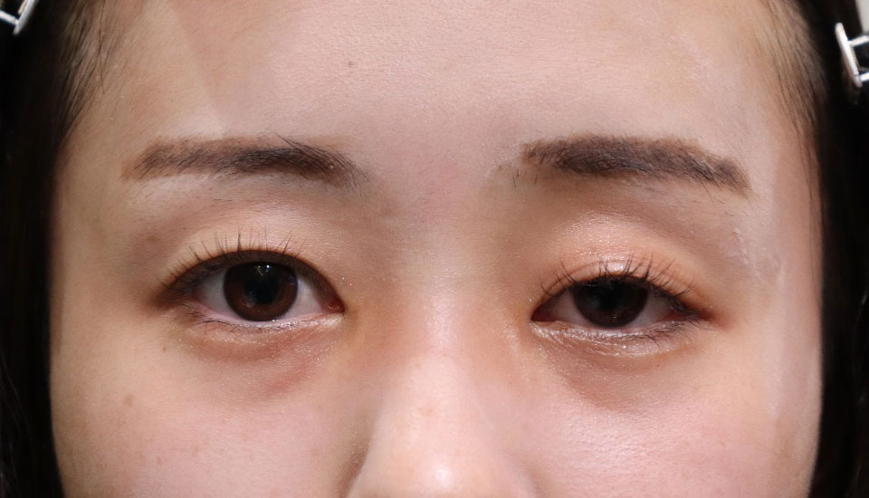
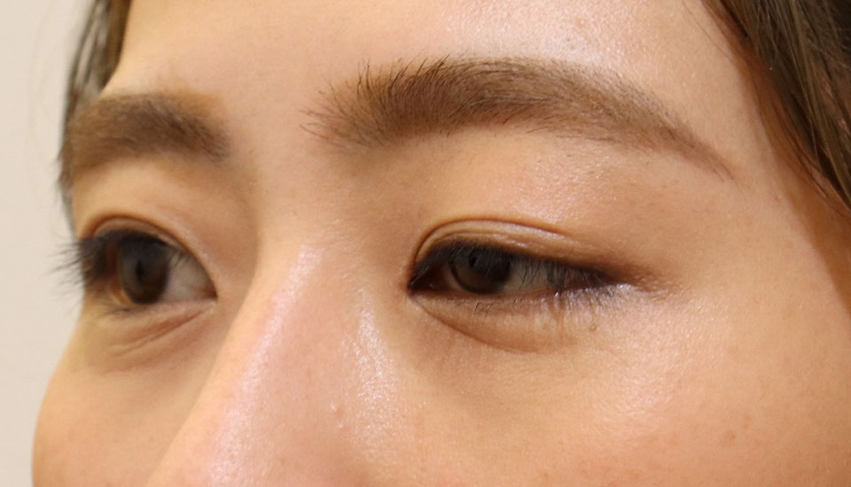
The condition is left congenital ptosis. The left has ptosis, and the right has skin laxity, resulting in a significant difference in the shape of the eyelids between the two sides.
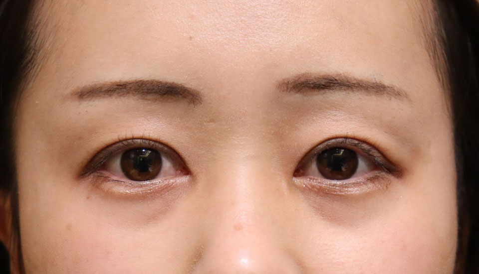
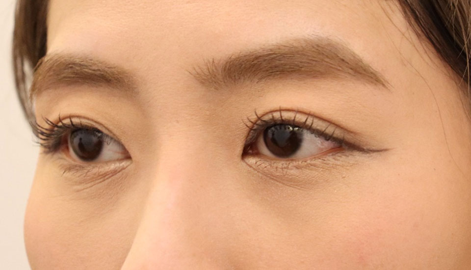
After the surgery, the width of the double eyelid and the degree of eye-opening are almost the same on both sides. The difference between the left and right is hardly noticeable now.
| Treatment details | Right eye:Excisional skin surgery+Double eyelid surgery Left eye:Levator muscle resection+Excisional skin surgery+Double eyelid surgery |
|---|---|
| Surgeon | Tomoyuki Kashima / MD, PhD |
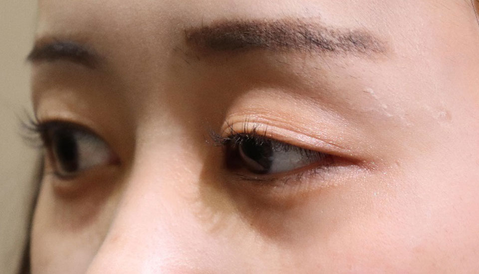
Her eyes have a somewhat dark impression due to ptosis.
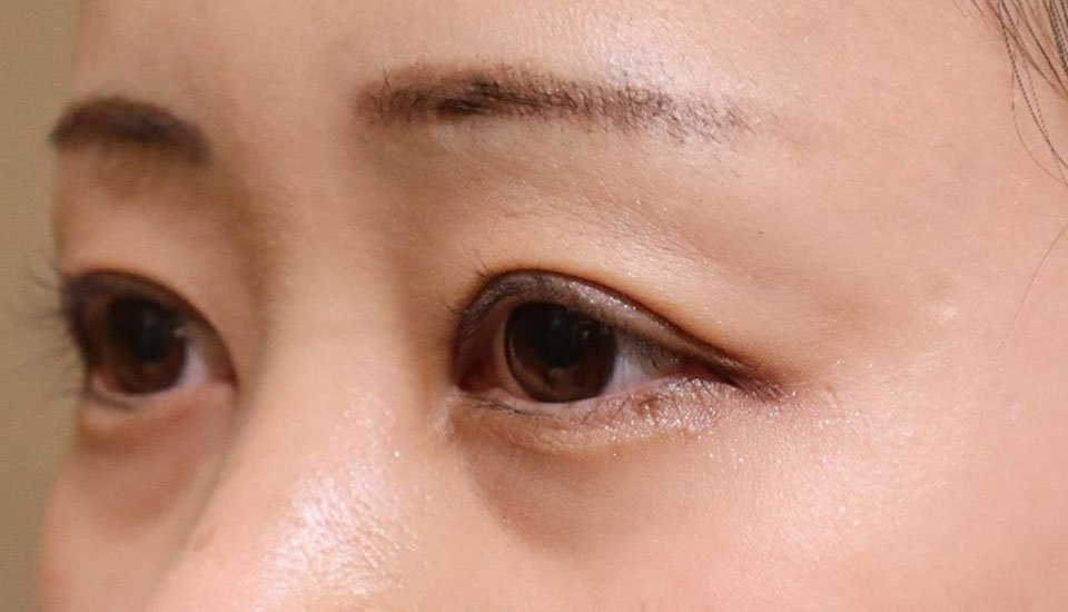
Just by curing the droopy eyelid, we were able to create this brighter impression.
30’s female.
She came to our hospital after one surgery at a university hospital and one surgery at a different large hospital for her left congenital ptosis without improvement.
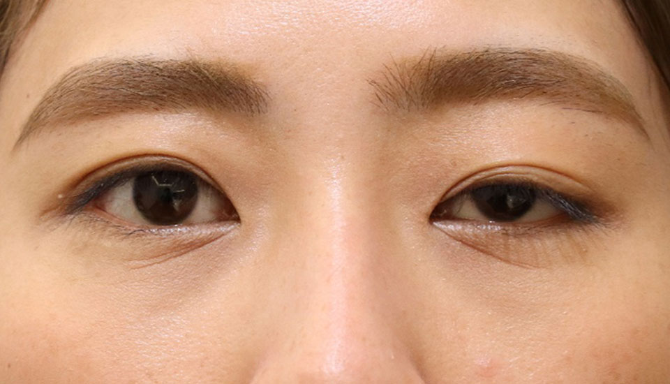
Due to a left congenital ptosis, the dark eye is underexposed and somewhat dark. 2 major hospitals performed surgery, but the elevation was inadequate.
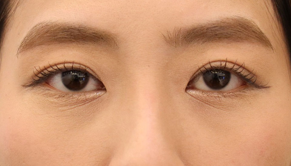
After the surgery, the width of the eyelid and the degree of widening of the eyelid are almost the same on the left and right sides. The difference between the left and right sides is almost imperceptible.
| Treatment details | Left eye:Levator muscle resection+Excisional skin surgery+Double eyelid surgery |
|---|---|
| Surgeon | Tomoyuki Kashima / MD, PhD |
Her eyelids are drooping, so she looks like she has a gloomy expression.
The asymmetry between the eyes has been corrected, and the double eyelid lines have become clean, making her eyes look very beautiful.

We perform over 5,000 ptosis surgeries annually, and as a result, we often take on cases that were unsuccessful at other institutions.
The outcomes of the surgeries vary by doctor, and in most cases, we are able to improve conditions even for patients whose previous surgeries at other institutions were unsuccessful. We encourage you not to give up and to consult with us.
Over the past 10 years, we have performed more than 10,000 eye plastic surgeries in Japan and the United States.
I aim to provide the cutting-edge medical knowledge and experience I have gained to patients in need in Japan.
| 2002 | Graduated from Gunma University Medical School Department of Ophthalmology |
|---|---|
| 2004 | Isesaki Municipal Hospital |
| 2005 | Gunma University Ophthalmology |
| 2007-09 | Seirei Hamamatsu Hospital Domestic study in Oculoplastic and Orbital Surgery |
| 2009 | Established an Oculoplastic Clinic at Gunma University |
| 2012 | Received degree Assistant Professor at Gunma University Department of Ophthalmology |
| 2010-18 | Director of the Asia-Pacific Society of Oculoplastic Surgery |
| 2015-16 | Studied at the University of California Los Angeles |
| 2017 | Opened Shin-Maebashi Kashima Oculoplastic Surgery Clinic |
| 2018 | Opened the Oculofacial Clinic in Tokyo |
| 2019 | Selected for The New York Times special feature "Next Era Leaders 2019" |
| 2020 | Member of the American Society of Ophthalmic Plastic and Reconstructive Surgery (ASOPRS) |
| 2009 | Singapore National Eye Center |
|---|---|
| 2010 | Asia-Pacific Society of Ophthalmic Plastic Surgery in Beijing Invited Speaker |
| 2011 | European Society of Ophthalmic Plastic Surgery in Como, Italy |
| 2012 |
World Ophthalmology Congress in Abu Dhabi Invited Speaker Asia ARVO Singapore Invited Speaker APAO Busan Invited Speaker American Academy of Ophthalmology Chicago Invited Speaker |
| 2013 | APAO Hyderabad Invited Speaker American Pediatric Ophthalmology Society Singapore Invited Speaker European Society of Ophthalmic Plastic Surgery Barcelona |
| 2014 | World Ophthalmology Congress Tokyo Invited Speaker Asia-Pacific Society of Ophthalmic Plastic Surgery Delhi American Society of Ophthalmic Plastic and Reconstructive Surgery Chicago |
| 2015 | APAO Guangzhou Invited Speaker |
| 2016 | KSAS (Korea Society of Aesthetic Surgery) meeting in Seoul ITEDS (International Thyroid Eye Disease) meeting in London AP SOPRS & JSOPRS joint meeting session chair iseminer Numerous lectures on surgical treatment of thyroid eye disease. |
| 2017 | American Academy of Ophthalmology instructor, Korean Society of Oculoplastic Surgery invited speaker, Chinese Society of Oculoplastic Surgery invited speaker |
| 2018 | Asia-Pacific International Society APAO invited speaker |
| 2019 | Asia-Pacific International Society APAO invited speaker, ITEDS invited speaker, OPAIC invited speaker |
| 2021 | Asia-Pacific Society of Ophthalmic Plastic Surgery invited speaker |

Perioperative care of the external eye
Meo Ice
A medical product developed by Nagoya Glasses to prevent "swelling" and "pain" after surgery around the eyelids.
Chief Editor
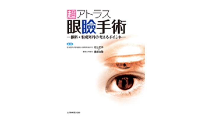
Ultimate Atlas of Eyelid Surgery
An ambitious atlas aiming for collaboration between ophthalmology and plastic surgery, published by All Japan Hospital Publishing. It is clearly explained with continuous color photos and detailed schematics.
Chief Editor
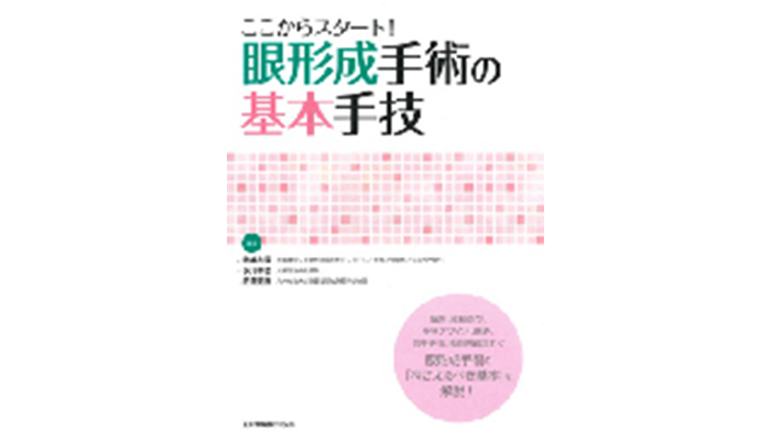
Start Here! Basic Techniques in Eyelid Plastic Surgery
An introductory book for doctors aspiring to specialize in oculoplastic surgery, published by All Japan Hospital Publishing. It thoroughly explains surgical tools and techniques.

| Office Hours | MON | TUE | WEN | THU | FRI | SAT | SUN |
|---|---|---|---|---|---|---|---|
| 8:15〜17:15 | - | ◯ | ◯ | ◯ | ◯ | ◯ | - |
[Closed]Monday & Sunday ,Japanese National Holiday.
Office Hours Calendar